REVIEW ARTICLE


Non-HLA Genes and Multiple Sclerosis


Jamilah Borjac1, #, Alaa Matar2, 3, #, Maxime Merheb4, Cijo George Vazhappilly4, Rachel Matar4, *
1 Department of Biological Sciences, Faculty of Science, Beirut Arab University, Debbieh, Lebanon
2 Department of Nursing, Faculty of Public Health, Jinan University, Tripoli, Lebanon
3 Department of Biological Sciences, Faculty of Science, Beirut Arab University, Tripoli, Lebanon
4 Department of Biotechnology, American University of Ras Al Khaimah AURAK, Ras Al Khaimah, P.O Box 10021, United Arab Emirates


Article Metrics

CrossRef Citations:
0
Total Statistics:

Full-Text HTML Views: 1598
Abstract HTML Views: 669
PDF Downloads: 554
ePub Downloads: 316
Total Views/Downloads: 3137
Unique Statistics:

Full-Text HTML Views: 879
Abstract HTML Views: 360
PDF Downloads: 403
ePub Downloads: 238
Total Views/Downloads: 1880


Creative Commons License
© 2023 Borjac et al.

open-access license: This is an open access article distributed under the terms of the Creative Commons Attribution 4.0 International Public License (CC-BY 4.0), a copy of which is available at: https://creativecommons.org/licenses/by/4.0/legalcode. This license permits unrestricted use, distribution, and reproduction in any medium, provided the original author and source are credited.

* Address Correspondence to this author at the Department of Biotechnology, American University of Ras Al Khaimah AURAK, Ras Al Khaimah, P.O Box 10021, United Arab Emirates; E-mail: rachel.matar@yahoo.fr
#These authors have contributed equally


Abstract

Multiple sclerosis (MS) is an autoimmune demyelinating disease of the central nervous system. Identification of genetic variants that pose risks to MS is of high interest since they contribute largely to disease pathogenesis. A rich body of literature associated these risks with variants of HLA genes located mostly on the short arm of chromosome 6 (6p21). These genetic variations may result in alteration in protein function and are associated, therefore, with disease phenotype and therapy outcome. Although the HLA region has been routinely known to have the strongest correlation with MS, other genes found within and outside HLA locus are considered risk factors for MS. The objective of this review is to shed light on the non-HLA genes implicated with multiple sclerosis. Due to the interplay between the polygenetic and environmental factors, along with their differential contribution and genetic heterogeneity among populations, it is extremely challenging to determine the contribution of the non-HLA genes to the outcome and onset of MS disease. We conclude that a better assemblage of genetic factors involved in MS can have a critical impact on the establishment of a genetic map of MS that allows proper investigation at the expression and functional levels.

Keywords: Multiple sclerosis, Non-HLA genes, Polymorphism, Autoimmune demyelinating disease, Genetic variants, CNS.


1. INTRODUCTION

Multiple sclerosis (MS) is an inflammatory neurodegenerative disorder of the central nervous system (CNS) resulting in a progressive loss of the myelin sheath that typically leads to sensory, motor, and neurocognitive disturbance [1]. According to the Atlas of MS, this unpredictable disease affects around 2.2 million people globally, under different clinical forms and manifestations [2]. Different data support the evidence that adults aged between 20 and 40 years are more susceptible to the disease, with a higher occurrence in females than males. Although MS etiology is poorly established, it is thought to be the consequence of environmental and genetic interactions [3]. Among environmental factors, smoking, infections, and sunlight exposure are the most widely reported [4-6]. Genetic factors related to MS are numerous and are believed to contribute to MS onset through various immunologically relevant genetic risk factors [7]. On one hand, HLA genes are believed to carry the strongest genetic risk variant for MS [8]. Consistently, it has been demonstrated that extended haplotypes spanning HLA class I and II are of high interest since they are implicated in the risk of several autoimmune diseases. On the other hand, non-HLA genes, including genes that may influence distinct immune tissues and cell types, contribute to the disease to a considerable extent. Recently, genome-wide associations studies performed by the International Multiple Sclerosis Genetics Consortium (IMSGC) analyzed genetic data from 47,429 MS patients and 68,374 individuals without MS in order to establish a genetic map for MS and have determined more than 230 independent MS risk variants, consisting of 200 autosomal variations, one chromosome X variation, and 32 different HLA ones. Various studies on different populations tackled the involvement of several non-HLA genes in MS with conflicting results. In this review, we will shed light on the mostly reported non-HLA genes along with their contribution to MS. These genes were chosen to be the major non-HLA contributors after a vigorous search conducted using Pubmed. In our search, we focused on related articles dated from January 2005 till January 2023.

2. NON-HLA GENES AND MS

Table 1 represents the main non-HLA genes that showed potential correlation with MS, along with their contribution to normal physiological conditions. These genes were the major non-HLA gene contributors obtained after a vigorous search conducted, using PubMed, from the published articles dated from January 2005 till January 2023.

Table 1. The main non-HLA genes show a potential correlation with MS along with their contribution to normal physiological conditions.
Non-HLA MS Susceptible Gene Role in Normal Physiological Condition References
CD58 Co-stimulation and proliferation of T-cell receptor signaling via its interaction with cluster of differentiation 2 (CD2) [9]
CD6 Co-stimulation and proliferation of T-cell receptor signaling via its interaction with the ligand activated leukocyte cell adhesion molecule (ALCAM) [10]
CLEC16A Retrograde transport of HLA-II containing compartments [11]
CYP27B1 Implication in converting vitamin D to its active form [12]
FoxP3 Control T-cell activity by inducing the activity of regulatory T (Treg) cells [13]
IL2-Rα Binding of interleukin 2 (IL2) resulting in the activation of T cells [14]
IL7-Rα Binding of interleukin 7 (IL7) resulting in the differentiation of T cells [15]

2.1. Cluster of Differentiation 58 (CD58)

CD58 encodes for a glycosylated cell adhesion molecule known as lymphocyte-associated antigen 3(LFA3) that is found on human chromosome 1p13. It is present on the surface of antigen-presenting cells (APCs), especially macrophages, and is able to promote their specific adherence to the CD2 ligand on the T-cell surface. A whole-genome association scan has proposed that genetic variations in CD58 are associated with MS risk. The most studied genetic variant is rs2300747, where inconclusive data were shown; an association was maintained in different studies [16-18] and supported by the fact that MS patients carrying the G protective allele presented higher CD58 mRNA expression level during clinical remission [17]. However, this genetic variant association with MS was not consistent among other studies [19, 20]. On the other hand, a second genetic variant rs12044852 was shown to be correlated with MS in Australians and Russians [18, 21] but not in Malay and Iranian [19, 22] populations where the CC genotype was strongly associated with multiple sclerosis severity score (MSSS), indicating by this a negative effect of this SNP in response to interferon-beta (IFN-β) therapy [22].

2.2. Cluster of Differentiation 6 (CD6)

CD6 encodes a cell surface scavenger implicated in thymocyte differentiation as well as in T-cell activation and differentiation. It has been suggested that CD6 may play a crucial role in MS pathogenesis as it was shown to be involved in the transmigration of leukocytes across the blood-brain barrier (BBB). However, its definite role in regulating T-cell responses remains controversial. Genome-wide association studies have identified a large number of genetic variants associated with autoimmune diseases, including MS. Previous reports showed an association between CD6 genetic variant rs17824933 and MS [23-25]. In addition, the risk allele of this single nucleotide polymorphism (SNP) was demonstrated to be associated with a decreased expression of full-length CD6 in CD4+ and CD8+ T cells, affecting consequently their proliferation [26]. Hence, targeting CD6 by developing anti-human CD6 monoclonal antibody would be of high interest in MS therapy as it prevents T cell depletion [27]. Additional risk variants were shown to be associated with MS like rs12360861 in Poland population and rs650258 in the Spanish-Basque population [28, 29] with data replication needed. Overall, these data reinforce the important role of CD6 in MS pathogenesis.

2.3. C-type Lectin Domain Containing 16A (CLEC16A)

C-type lectins are key players in immune regulation as they drive different functions of antigen presenting cells (APCs) [30]. Located on chromosome 16p13, a susceptible locus for various autoimmune diseases, this gene is considered among the first non-HLA genes associated with MS [21, 31-33]. Moreover, upregulation of CLEC16A was observed in T cells of MS patients homozygous for the risk allele rs12927355 CLEC16A [34]. Additionally, higher expression of CLEC16A was detected in the white matter of MS patients, especially in the peripheral blood mononuclear cells (PBMCs). The high expression of this gene was blunted when patients were treated with vitamin D, indicating by this that CLEC16A may play a pivotal role in MS pathogenesis [11]. Additional functional studies are required to better understand the role of CLEC16A in MS as well as in other autoimmune diseases.

2.4. Cytochrome P450 Family 27 Superfamily B Peptide 1 (CYP27B1)

CYP27B1, located on chromosome 12q13-14, encodes for vitamin D metabolizing enzyme, the hydroxyvitamin D3-1-alpha-hydroxylase. Pre-vitamin D3 is produced in the skin and converted to 25(OH)D3 in the liver. In skin, kidney and immune cells, CYP27B1 enzyme converts 25(OH)D3 into 1,25 (OH)2D3 that binds to the vitamin D receptor present at the surface of T-cells and antigen presenting cells (APCs). Consequently, it suppresses the adaptive immune response, decreases dendritic cell and T-cell proliferation, differentiation, and maturation as well as Th1/Th2 ratio, and enhances the suppressive function of regulatory T-cells. Several studies have highlighted the role of rs703842 in MS with inconsistent results reported. Most studies showed an association between this risk variant and MS in Caucasian [35, 36], Slovakian [37], and Han Chinese [12] populations but not in others [38-40]. This was associated with a lower level of vitamin D registered in MS patients compared with controls, regardless of the risky genotypic and allelic status of this variant [35, 38]. One additional risk variant, rs118204009 was suggested to be implicated in MS [12, 41]. Further consideration of distinct ethnicity groups is needed for a better understanding of CYP27B1 role in MS.

2.5. Forkhead box P3 (FoxP3)

FoxP3 encodes a transcription factor that is predominantly expressed in CD4(+) CD25(+) regulatory T cells, playing a key role in maintaining immune homeostasis. It is considered the master transcription factor of these cells, responsible for the polarization of naïve T cells into Treg cells. Recently, T regulatory (Treg) cells have been known to present an impaired suppressive function in MS disease [42]. Accumulating evidence showed that functional alterations in FoxP3 gene expression have been observed in several autoimmune diseases [43, 44], linking thereby the defect in functional peripheral immunomodulation to an established genetic variant implicated in immune regulation and autoimmunity. A positive correlation was found between the genetic variant rs3761548 and MS [44-48]. Additional single nucleotide polymorphisms were also investigated with conflicting results [47, 50]. FoxP3 gene expression level was validated to be decreased in experimental autoimmune encephalomyelitis (EAE) models or in MS patients [51-53], which could not be reproduced in the study of Akbari et al. [54]. Interestingly, FoxP3 expression level could be restored by (IFN-β) treatment [55] or through injection of ex vivo autologous Treg cells that helped in increasing the level of Treg in patients’ blood. To sum up, FoxP3 could share a powerful link to MS susceptibility as it exerts an immunomodulatory effect.

Fig. (1). This figure represents a functional analysis of the main non-HLA MS susceptible genes and their potential contribution to MS. The risk allele rs703842 of CYP27B1 induces a reduced production of IL2. Those of CD6 (rs17824933 and rs12360861) induce a decrease in CD6 expression and that of IL7-Rα (rs6897932) leads to the production of the soluble form of IL7-Rα. All four SNPs (rs703842, rs17824933, rs12360861, and rs6897932) result in the alteration of T cell proliferation that could affect MS susceptibility. The risk allele rs12044852 of CD58 induces a decrease in CD58 expression that leads to FoxP3 down-regulation. FoxP3 expression may also be down-regulated due to the risk allele rs3761548 of FoxP3. The reduced expression of FoxP3 ends up with the dysfunction of regulatory T (Treg) cells that will increase the risk for MS susceptibility. On the contrary, the protective allele (rs2300747) of CD58 induces an increase in CD58 expression that will lead to FoxP3 up-regulation and consequently to Treg cell activation, reducing the risk for MS susceptibility. The numbers in brackets above each box represent the references used in linking the main non-HLA gene SNPs studied to their gene expression level and their potential contribution to MS.

2.6. Interleukin 2- receptor Alpha (IL2-Rα)

IL2-Rα, also known as CD25, is located on chromosome 10 and encodes the specific component of the high affinity IL2-R system that is implicated in autoimmunity and immune-regulation, where IL2/IL2-R signaling pathway allows the proliferation and survival of affected T cells and regulatory T cells production [56, 57]. Genome wide-association studies and fine mapping have revealed a tight link between SNPs in IL2-Rα and increased risk of immune mediated diseases including MS. Different studies were conducted on IL2-Rα polymorphisms and MS, mostly studying SNP rs2104286 that showed a risk susceptibility for the disease according to several publications [21, 58-61]. Additionally, one report has found that this SNP was accompanied by a reduced frequency of CD25(+) follicular helper T1 (TFH1) cells in patients carrying the risk genotype [62]. Fewer studies were performed on the rs12722489 SNP that also showed to be implicated with MS [59, 63]. However, data replication is needed on various populations for further validation. Furthermore, a study by Ainiding et al. showed that DNA hypo-methylation of IL2-Rα was significantly associated with MS and was accompanied by a higher expression of IL2-Rα in T cells of MS patients [64]. Together, these data confirm the tight correlation between IL2-Rα and MS.

2.7. Interleukin 7- receptor Alpha (IL7-Rα)

IL7-Rα, located on 5p13 human chromosome, encodes a subunit of IL7 receptor that plays a role in immune homeostasis by assisting in the maturation of B and T cells. Various genome wide association studies revealed that IL7-Rα is correlated with various immunological disorders [65], such as MS [66], and thus is considered among the top listed candidate genes implicated in MS. Evidence illustrates the tight association between MS and SNPs in the promoter and exon region of IL7-Rα. Genotyping of 123 SNPs in 66 genes chosen according to their chromosomal location or biological roles has identified that IL7-R includes at least 3 significantly associated SNPs with MS risk [67]. Additionally, alteration in the expression of genes encoding IL7-Rα and its IL7 ligand has been shown in the cerebrospinal fluid (CSF) compartment of MS patients [68]. Most studies tackled the association between genetic variant located in exon 6 of IL7-Rα and MS with a well-established association [63, 69-77] or association reach-ing significance after stratification analysis in progressive MS subjects only [78, 79] or more specifically in secondary- progressive MS (SPMS) [80]. However, no association was found in other studies [81-83]. It has been suggested that rs6897932 risk variant is linked to altered alternative splicing of exon 6 that contributes to its skipping, affecting therefore, the ratios of soluble (sIL7- Rα) to membrane-bound IL7-Rα [84]. However, a reduced expression of sIL7-Rα was detected in progressive MS subjects regardless of their genotypes [78] and showed no effect of genotype or protein isoforms expression on MS phenotype [71]. Furthermore, a genetic variant in the 5’UTR of the RNA helicase DDX39B, a potent activator of exon 6 of IL7-Rα, was shown to reduce its translation, resulting in increased levels of the soluble form of IL7-Rα [85-88]. A meta-analysis study consisting of 9734 cases and 10436 controls confirmed the association between 3 SNPs of IL7-Rα (rs3194051 in exon 8, rs987107 in the intronic region, and rs11567686 in the promoter) and MS [66]. To sum up, IL7-Rα locus polymorphisms can have a key role in MS predisposition.

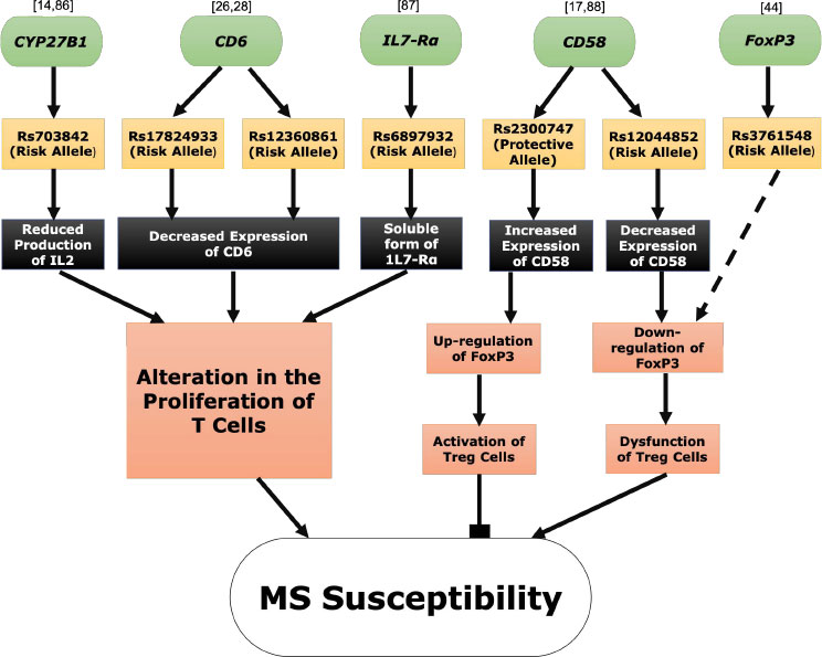
Fig. (1) depicts a functional analysis of the main non-HLA MS susceptible genes and their potential contribution to MS.

CONCLUSION

Multiple sclerosis is a chronic autoimmune disorder where genetic variations, especially those involved with immune regulation, play a key role in its development. The literature on HLA related genes in MS is rich, including genetic and functional studies. Importantly, the latter showed that HLA genetic associations with MS were linked to functions not directly associated with antigen presentation. On the other hand, although non-HLA genetic variations were prominently associated with MS, conflicting results were reported based on the genetic backgrounds. Thus, more studies are needed to verify the functional impact of these variants in MS progression. In this review, we summarized the most relevant non-HLA genetic variants associated with MS. Overall, these variants may serve as a starting point for MS genetic map construction for a further investigation of these genes at transcriptional, functional, and signaling pathway levels.

LIST OF ABBREVIATIONS

MS = Multiple Sclerosis
CNS = Central Nervous System
IMSGC = International Multiple Sclerosis Genetics Consortium

CONSENT FOR PUBLICATION

Not applicable.

FUNDING

None.

CONFLICT OF INTEREST

The authors declare no conflict of interest financial or otherwise.

ACKNOWLEDGEMENTS

Declared none.

REFERENCES

[1] Feinstein A, Brochet B, Sumowski J. The cognitive effects of anxiety and depression in immune-mediated inflammatory diseases. Neurology 2019; 92(5): 211-2.
[2] Wallin MT, Culpepper WJ, Nichols E, et al. Global, regional, and national burden of multiple sclerosis 1990–2016: A systematic analysis for the Global Burden of Disease Study 2016. Lancet Neurol 2019; 18(3): 269-85.
[3] Olsson T, Barcellos LF, Alfredsson L. Interactions between genetic, lifestyle and environmental risk factors for multiple sclerosis. Nat Rev Neurol 2017; 13(1): 25-36.
[4] Nishanth K, Tariq E, Nzvere FP, Miqdad M, Cancarevic I. Role of smoking in the pathogenesis of multiple sclerosis: A review article. Cureus 2020; 12(8): e9564.
[5] Castelo-Branco A, Chiesa F, Conte S, et al. Infections in patients with multiple sclerosis: A national cohort study in Sweden. Mult Scler Relat Disord 2020; 45: 102420.
[6] Espinosa-Ramírez G, Ordoñez G, Flores-Rivera J, Sotelo J. Sunlight exposure and multiple sclerosis in a tropical country. Neurol Res 2014; 36(7): 647-50.
[7] Navaderi M, Rajaei S, Rahimirad S, et al. Identification of Multiple Sclerosis key genetic factors through multi-staged data mining. Mult Scler Relat Disord 2020; 39: 101446.
[8] Alcina A, Abad-Grau MM, Fedetz M, et al. Multiple sclerosis risk variant HLA-DRB1*1501 associates with high expression of DRB1 gene in different human populations. PLoS One 2012; 7(1): e29819.
[9] Davis SJ, van der Merwe PA. The structure and ligand interactions of CD2: Implications for T-cell function. Immunol Today 1996; 17(4): 177-87.
[10] Zimmerman AW, Joosten B, Torensma R, Parnes JR, van Leeuwen FN, Figdor CG. Long-term engagement of CD6 and ALCAM is essential for T-cell proliferation induced by dendritic cells. Blood 2006; 107(8): 3212-20.
[11] van Luijn MM, Kreft KL, Jongsma ML, et al. Multiple sclerosis-associated CLEC16A controls HLA class II expression via late endosome biogenesis. Brain 2015; 138(6): 1531-47.
[12] Zhuang JC, Huang ZY, Zhao GX, Yu H, Li ZX, Wu ZY. Variants of CYP27B1 are associated with both multiple sclerosis and neuromyelitis optica patients in Han Chinese population. Gene 2015; 557(2): 236-9.
[13] Alessandra C, Fortunata C, Sara B, et al. Molecular mechanisms controlling foxp3 expression in health and autoimmunity: From epigenetic to post-translational regulation. Front Immunol 2020; 10: 3136.
[14] Kendall A. Interleukin-2. Curr Opin Immunol 1992; 4(3): 271-6.
[15] Chazen GD, Pereira GM, LeGros G, Gillis S, Shevach EM. Interleukin 7 is a T-cell growth factor. Proc Natl Acad Sci USA 1989; 86(15): 5923-7.
[16] Liu J, Liu X, Liu Y, et al. Association of EVI5 rs11808092, CD58 rs2300747, and CIITA rs3087456 polymorphisms with multiple sclerosis risk: A meta-analysis. Meta Gene 2016; 9: 97-103.
[17] De Jager PL, Baecher-Allan C, Maier LM, et al. The role of the CD58 locus in multiple sclerosis. Proc Natl Acad Sci USA 2009; 106(13): 5264-9.
[18] Lezhnyova V, Davidyuk Y, Mullakhmetova A, et al. Analysis of herpesvirus infection and genome single nucleotide polymorphism risk factors in multiple sclerosis, Volga federal district, Russia. Front Immunol 2022; 13: 1010605.
[19] Ching YM, Viswanathan S, Mohamed Nor N, et al. Association of CD58 polymorphism and multiple sclerosis in Malaysia: A pilot study. Auto Immun Highlights 2019; 10(1): 13.
[20] Ghavimi R, Alsahebfosoul F, Salehi R, Kazemi M, Etemadifar M, Zavaran Hosseini A. High-resolution melting curve analysis of polymorphisms within CD58, CD226, HLA-G genes and association with multiple sclerosis susceptibility in a subset of Iranian population: A case–control study. Acta Neurol Belg 2020; 120(3): 645-52.
[21] Rubio JP, Stankovich J, Field J, et al. Replication of KIAA0350, IL2RA, RPL5 and CD58 as multiple sclerosis susceptibility genes in Australians. Genes Immun 2008; 9(7): 624-30.
[22] Torbati S, Karami F, Ghaffarpour M, Zamani M. Association of CD58 polymorphism with multiple sclerosis and response to interferon ß therapy in a subset of Iranian Population. Cell J 2015; 16(4): 506-13.
[23] D’Cunha MA, Pandit L, Malli C. CD6 gene polymorphism rs17824933 is associated with multiple sclerosis in Indian population. Ann Indian Acad Neurol 2016; 19(4): 491-4.
[24] Chatenoud L, Ed. The genetic association of variants in CD6, TNFRSF1A and IRF8 to multiple sclerosis: A multicenter case-control study. PLoS One 2011; 6(4): e18813.
[25] Swaminathan B, Matesanz F, Cavanillas ML, et al. Validation of the CD6 and TNFRSF1A loci as risk factors for multiple sclerosis in Spain. J Neuroimmunol 2010; 223(1-2): 100-3.
[26] Kofler DM, Severson CA, Mousissian N, De Jager PL, Hafler DA. The CD6 multiple sclerosis susceptibility allele is associated with alterations in CD4+ T cell proliferation. J Immunol 2011; 187(6): 3286-91.
[27] Li Y, Singer NG, Whitbred J, Bowen MA, Fox DA, Lin F. CD6 as a potential target for treating multiple sclerosis. Proc Natl Acad Sci USA 2017; 114(10): 2687-92.
[28] Wagner M, Bilinska M, Pokryszko-Dragan A, et al. ALCAM and CD6 — multiple sclerosis risk factors. J Neuroimmunol 2014; 276(1-2): 98-103.
[29] Swaminathan B, Cuapio A, Alloza I, et al. Fine mapping and functional analysis of the multiple sclerosis risk gene CD6. PLoS One 2013; 8(4): e62376.
[30] Guasconi L, Serradell MC, Garro AP, Iacobelli L, Masih DT. C-type lectins on macrophages participate in the immunomodulatory response to Fasciola hepatica products. Immunology 2011; 133(3): 386-96.
[31] Nischwitz S, Cepok S, Kroner A, et al. More CLEC16A gene variants associated with multiple sclerosis. Acta Neurol Scand 2011; 123(6): 400-6.
[32] Hoppenbrouwers IA, Aulchenko YS, Janssens AC, et al. Replication of CD58 and CLEC16A as genome-wide significant risk genes for multiple sclerosis. J Hum Genet 2009; 54(11): 676-80.
[33] Zoledziewska M, Costa G, Pitzalis M, et al. Variation within the CLEC16A gene shows consistent disease association with both multiple sclerosis and type 1 diabetes in Sardinia. Genes Immun 2009; 10(1): 15-7.
[34] Leikfoss IS, Keshari PK, Gustavsen MW, et al. Multiple sclerosis risk allele in CLEC16A acts as an expression quantitative trait locus for CLEC16A and SOCS1 in CD4+ T Cells. PLoS One 2015; 10(7): e0132957.
[35] Smagina IV, Lunev KV, Elchaninova SA. Association between vitamin D status and CYP27b1 and CYP24A1 gene polymorphisms in patients with multiple sclerosis in the Altai region. Zh Nevrol Psikhiatr Im S S Korsakova 2020; 120(2): 61-6.
[36] Jiang T, Li L, Wang Y, et al. The association between genetic polymorphism rs703842 in CYP27B1 and multiple sclerosis. Medicine 2016; 95(19): e3612.
[37] Čierny D, Michalik J, Dubovan P, et al. The association of rs703842 variants in CYP27B1 with multiple sclerosis susceptibility is influenced by the HLA-DRB1*15:01 allele in Slovaks. J Neuroimmunol 2019; 330: 123-9.
[38] Agnello L, Scazzone C, Lo Sasso B, et al. VDBP, CYP27B1, and 25-Hydroxyvitamin D gene polymorphism analyses in a group of sicilian multiple sclerosis patients. Biochem Genet 2017; 55(2): 183-92.
[39] Barizzone N, Pauwels I, Luciano B, et al. No evidence for a role of rare CYP27B1 functional variations in multiple sclerosis. Ann Neurol 2013; 73(3): 433-7.
[40] Ban M, Caillier S, Mero IL, et al. No evidence of association between mutant alleles of the CYP27B1 gene and multiple sclerosis. Ann Neurol 2013; 73(3): 430-2.
[41] Ross JP, Bernales CQ, Lee JD, Sadovnick AD, Traboulsee AL, Vilariño-Güell C. Analysis of CYP27B1 in multiple sclerosis. J Neuroimmunol 2014; 266(1-2): 64-6.
[42] Mexhitaj I, Nyirenda MH, Li R, et al. Abnormal effector and regulatory T cell subsets in paediatric-onset multiple sclerosis. Brain 2019; 142(3): 617-32.
[43] Wu Y, Zhao Y, Xu T, You L, Zhang H, Liu F. Alzheimer’s disease affects severity of asthma through methylation control of Foxp3 promoter. J Alzheimers Dis 2019; 70(1): 121-9.
[44] Zhang Y, Zhang J, Liu H, et al. Meta-analysis of FOXP3 gene rs3761548 and rs2232365 polymorphism and multiple sclerosis susceptibility. Medicine 2019; 98(38): e17224.
[45] Flauzino T, Alfieri DF, de Carvalho Jennings Pereira WL, et al. The rs3761548 FOXP3 variant is associated with multiple sclerosis and transforming growth factor β1 levels in female patients. Inflamm Res 2019; 68(11): 933-43.
[46] Wawrusiewicz-Kurylonek N, Chorąży M, Posmyk R, et al. The FOXP3 rs3761547 gene polymorphism in multiple sclerosis as a male-specific risk factor. Neuromolecular Med 2018; 20(4): 537-43.
[47] Eftekharian MM, Sayad A, Omrani MD, et al. Single nucleotide polymorphisms in the FOXP3 gene are associated with increased risk of relapsing-remitting multiple sclerosis. Hum Antibodies 2017; 24(3-4): 85-90.
[48] Jafarzadeh A, Jamali M, Mahdavi R, et al. Circulating levels of interleukin-35 in patients with multiple sclerosis: Evaluation of the influences of FOXP3 gene polymorphism and treatment program. J Mol Neurosci 2015; 55(4): 891-7.
[49] Kamal A, Hosny M, Abd Elwahab A, Shawki Kamal Y, Shehata HS, Hassan A. FOXP3 rs3761548 gene variant and interleukin-35 serum levels as biomarkers in patients with multiple sclerosis. Rev Neurol (Paris) 2021; 177(6): 647-54.
[50] Işik N, Yildiz Manukyan N, Aydin Cantürk İ, Candan F, Ünsal Çakmak A, Saru Han Direskeneli G. Genetic susceptibility to multiple sclerosis: The role of FOXP3 gene polymorphism. Noro Psikiyatri Arsivi 2014; 51(1): 69-73.
[51] Etesam Z, Nemati M, Ebrahimizadeh MA, et al. Altered expression of specific transcription factors of Th17 (RORγt, RORα) and Treg Lymphocytes (FOXP3) by peripheral blood mononuclear cells from patients with multiple sclerosis. J Mol Neurosci 2016; 60(1): 94-101.
[52] Huan J, Culbertson N, Spencer L, et al. Decreased FOXP3 levels in multiple sclerosis patients. J Neurosci Res 2005; 81(1): 45-52.
[53] Mohajeri M, Farazmand A, Mohyeddin Bonab M, Nikbin B, Minagar A. FOXP3 gene expression in multiple sclerosis patients pre- and post mesenchymal stem cell therapy. Iran J Allergy Asthma Immunol 2011; 10(3): 155-61.
[54] Akbari Z, Taheri M, Jafari A, Sayad A. FOXP3 gene expression in the blood of Iranian multiple sclerosis patients. Hum Antibodies 2018; 26(3): 159-64.
[55] Vandenbark AA, Huan J, Agotsch M, et al. Interferon-beta-1a treatment increases CD56bright natural killer cells and CD4+CD25+ Foxp3 expression in subjects with multiple sclerosis. J Neuroimmunol 2009; 215(1-2): 125-8.
[56] Lifshitz GV, Zhdanov DD, Lokhonina AV, et al. Ex vivo expanded regulatory T cells CD4 + CD25 + FoxP3 + CD127 Low develop strong immunosuppressive activity in patients with remitting-relapsing multiple sclerosis. Autoimmunity 2016; 49(6): 388-96.
[57] Eliseeva DD, Lifshitz GV, Lokhonina AV, Zhdanov DD, Zavalishin IA, Bykovskaia SN. The treatment by expanded ex vivo autologous regulatory T-cells CD4+CD25+FoxP3+CD127low restores the balance of immune system in patients with remitting-relapsing multiple sclerosis. Zh Nevrol Psikhiatr Im S S Korsakova 2016; 116(2. Vyp. 2): 54-62.
[58] Stefanović M, Životić I, Stojković L, Dinčić E, Stanković A, Živković M. The association of genetic variants IL2RA rs2104286, IFI30 rs11554159 and IKZF3 rs12946510 with multiple sclerosis onset and severity in patients from Serbia. J Neuroimmunol 2020; 347: 577346.
[59] Xia ZL, Qin QM, Zhao QY. A genetic link between CXCR5 and IL2RA gene polymorphisms and susceptibility to multiple sclerosis. Neurol Res 2018; 40(12): 1040-7.
[60] Wang XX, Chen T. Meta-analysis of the association of IL2RA polymorphisms rs2104286 and rs12722489 with multiple sclerosis risk. Immunol Invest 2018; 47(5): 431-42.
[61] Alcina A, Fedetz M, Ndagire D, et al. IL2RA/CD25 gene polymorphisms: Uneven association with multiple sclerosis (MS) and type 1 diabetes (T1D). PLoS One 2009; 4(1): e4137.
[62] Buhelt S, Søndergaard HB, Oturai A, Ullum H, von Essen MR, Sellebjerg F. Relationship between multiple sclerosis-associated IL2RA risk allele variants and circulating T cell phenotypes in healthy genotype-selected controls. Cells 2019; 8(6): 634.
[63] Weber F, Fontaine B, Cournu-Rebeix I, et al. IL2RA and IL7RA genes confer susceptibility for multiple sclerosis in two independent European populations. Genes Immun 2008; 9(3): 259-63.
[64] Ainiding G, Kawano Y, Sato S, et al. Interleukin 2 receptor α chain gene polymorphisms and risks of multiple sclerosis and neuromyelitis optica in southern Japanese. J Neurol Sci 2014; 337(1-2): 147-50.
[65] Lundtoft C, Seyfarth J, Jacobsen M. IL7RA genetic variants differentially affect IL-7Rα expression and alternative splicing: A role in autoimmune and infectious diseases? Genes Immun 2020; 21(2): 83-90.
[66] Liu H, Huang J, Dou M, et al. Variants in the IL7RA gene confer susceptibility to multiple sclerosis in Caucasians: Evidence based on 9734 cases and 10436 controls. Sci Rep 2017; 7(1): 1207.
[67] Zhang Z, Duvefelt K, Svensson F, et al. Two genes encoding immune-regulatory molecules (LAG3 and IL7R) confer susceptibility to multiple sclerosis. Genes Immun 2005; 6(2): 145-52.
[68] Lundmark F, Duvefelt K, Iacobaeus E, et al. Variation in interleukin 7 receptor α chain (IL7R) influences risk of multiple sclerosis. Nat Genet 2007; 39(9): 1108-13.
[69] Gregory SG, Schmidt S, Seth P, et al. Interleukin 7 receptor α chain ( IL7R ) shows allelic and functional association with multiple sclerosis. Nat Genet 2007; 39(9): 1083-91.
[70] Zhang R, Duan L, Jiang Y, et al. Association between the IL7R T244I polymorphism and multiple sclerosis: A meta-analysis. Mol Biol Rep 2011; 38(8): 5079-84.
[71] Sombekke MH, van der Voort LF, Kragt JJ, et al. Relevance of IL7R genotype and mRNA expression in Dutch patients with multiple sclerosis. Mult Scler 2011; 17(8): 922-30.
[72] Taheri M, Sayad A. Investigating the exon 6 sequence changes of interleukin 7 receptor A (IL7RA) gene in patients with relapsing-remitting multiple sclerosis. Hum Antibodies 2019; 26(2): 43-8.
[73] Sahami-Fard MH, Mozhdeh M, Izadpanah F, Kashani HH, Nezhadi A. Interleukin 7 receptor T244I polymorphism and the multiple sclerosis susceptibility: A meta-analysis. J Neuroimmunol 2020; 341: 577166.
[74] AL-Eitan L, Al Qudah M, Al Qawasmeh M. Candidate gene association analysis of multiple sclerosis in the Jordanian Arab population: A case-control study. Gene 2020; 758: 144959.
[75] Razavian T, Shakib ME, Gharagozli K, et al. Association of rs12487066, rs12044852, rs10735781, rs3135388, rs6897932, rs1321172, rs10492972, and rs9657904 polymorphisms with multiple sclerosis in Iranian Population. Oman Med J 2020; 35(4): e150.
[76] Omraninava M, Mehranfar S, Vahedi P, et al. Association between IL7 Receptor Alpha (Il7ra) gene rs6897932 polymorphism and the risk of Multiple Sclerosis: A meta-regression and meta-analysis. Mult Scler Relat Disord 2021; 48: 102687.
[77] Zhao Z, Xue J, Zhuo Z, Zhong W, Liu H. The Association of IL7R rs6897932 with Risk of Multiple Sclerosis in Southern Chinese. Neuropsychiatr Dis Treat 2022; 18: 1855-9.
[78] Akkad DA, Hoffjan S, Petrasch-Parwez E, Beygo J, Gold R, Epplen JT. Variation in the IL7RA and IL2RA genes in German multiple sclerosis patients. J Autoimmun 2009; 32(2): 110-5.
[79] Traboulsee AL, Bernales CQ, Ross JP, Lee JD, Sadovnick AD, Vilariño-Güell C. Genetic variants in IL2RA and IL7R affect multiple sclerosis disease risk and progression. Neurogenetics 2014; 15(3): 165-9.
[80] Heidari M, Behmanesh M, Sahraian MA. Variation in SNPs of the IL7Ra gene is associated with multiple sclerosis in the Iranian population. Immunol Invest 2011; 40(3): 279-89.
[81] Vinoy N, Sheeja N, Kumar S, Biswas L. Class II HLA (DRB1, & DQB1) alleles and IL7R (rs6897932) variants and the risk for multiple sclerosis in Kerala, India. Mult Scler Relat Disord 2021; 50: 102848.
[82] Ünsal MA, Manukyan NY, IŞik N, Dİreskenelİ GS. Assessment of IL-7RA T244I polymorphism as a risk factor of multiple sclerosis in Turkish population. Noro Psikiyatri Arsivi 2019; 57(4): 280-2.
[83] Matar A, Jennani S, Abdallah H, Borjac J. Association of various genes with susceptibility to multiple sclerosis in Lebanese population of Bekaa region: A preliminary study. Kragujevac J Sci 2020; 42(42): 97-112.
[84] Evsyukova I, Bradrick SS, Gregory SG, Garcia-Blanco MA. Cleavage and polyadenylation specificity factor 1 (CPSF1) regulates alternative splicing of interleukin 7 receptor (IL7R) exon 6. RNA 2013; 19(1): 103-15.
[85] Galarza-Muñoz G, Briggs FBS, Evsyukova I, et al. Human epistatic interaction controls IL7R splicing and increases multiple sclerosis risk. Cell 2017; 169(1): 72-84.e13.
[86] Lu M, Shi H, Taylor BV, Körner H. Alterations of subset and cytokine profile of peripheral T helper cells in PBMCs from Multiple Sclerosis patients or from individuals with MS risk SNPs near genes CYP27B1 and CYP24A1. Cytokine 2022; 153: 155866.
[87] Alcina A, Fedetz M, Ndagire D, et al. The T244I variant of the interleukin-7 receptor-alpha gene and multiple sclerosis. Tissue Antigens 2008; 72(2): 158-61.
[88] Arthur AT, Armati PJ, Bye C, et al. Genes implicated in multiple sclerosis pathogenesis from consilience of genotyping and expression profiles in relapse and remission. BMC Med Genet 2008; 9(1): 17.