All published articles of this journal are available on ScienceDirect.

Comprehensive Genotypic Characterization of Antiseptic Resistance Determinants in Pseudomonas aeruginosa Clinical Isolates

Abstract
Introduction
The growing application of biocides post-COVID-19 has heightened concern over the development and spread of antiseptic resistance determinants, especially qac genes in different types of bacteria. This study aimed to evaluate antiseptic resistance determinants in clinical P. aeruginosa isolates and to determine their response to antimicrobial resistance and biofilm-forming ability.
Methods
Fifty clinical P. aeruginosa isolates were evaluated for antimicrobial susceptibility using the Kirby-Bauer disk diffusion method, biofilm formation using the microtiter plate crystal violet assay, and biocide tolerance by broth microdilution. PCR was performed to identify qacE and qacEΔ1 genes in selected highly resistant isolates. Statistical analysis was applied to identify the correlation among variables.
Results
The findings showed a high rate of MDR (64.0%) and XDR (6.0%). Biofilm assays showed strong (38.0%), moderate (30.0%), and weak (30.0%) production. Furthermore, strong /moderate biofilm formation was significantly associated with MDR/XDR phenotypes (p < 0.0001). Reduced antiseptic sensitivity was detected in (60.0%) of isolates and strongly correlated with both antimicrobial resistance status and biofilm strength (p < 0.0001). Molecular profiling of selected resistance isolates showed a universal presence of the qacEΔ1 gene (100%) and high prevalence of the qacE gene (73.3%).
Discussion
The significant correlation between qac genes and strong biofilm ability demonstrates a key mechanism of antibiotic and biocide tolerance in P. aeruginosa.
Conclusion
Qac genes are highly prevalent in clinical P. aeruginosa isolates and strongly correlated with multidrug resistance and biofilm-forming ability. Ongoing molecular monitoring of the biocide resistance genes in clinical setups is vital to prevent the propagation of exceptionally stubborn strains in clinical environments.
1. INTRODUCTION
Pseudomonas aeruginosa is a very versatile opportunistic pathogen that causes a broad range of healthcare-associated infections, especially among immunocompromised patients and those with long-term hospitalization or invasive interventions [1, 2]. Its resistance, metabolic plasticity, and adaptation to live in nutrient-limited environments allow it to survive in a great variety of ecological and clinical environments such as water systems, respiratory apparatus, wounds, and intensive care units. The extreme ability of the organism to survive under the influence of antiseptics and biocides also complicates the process of infection control, particularly in high-risk hospital settings where biocides are still needed to decontaminate the surfaces and instruments [3, 4].
P. aeruginosa exhibits strong biocide resistance with a close association with acquisition and transmission of qac (quaternary ammonium compound) genes, both qacE and a truncated form of qacE, qacEΔ1 [5]. These genes are typically incorporated in class 1 integrons and encode membrane transport proteins, which promote active efflux of quaternary ammonium compounds, chlorhexidine, benzalkonium chloride, and other antiseptics. Although the qacE variants are partially deleted, the qacEΔ1 variant still has the functional ability that reduces susceptibility to various antiseptic agents as well as increases environmental stability of resistant strains [6-8]. The qacE and qacEΔ1 determinants are also of great concern because of the integrase-mediated mobility that allows them to co-evolve with antibiotic resistance genes and thus allows the co-selection of multidrug-resistant (MDR) and extensively drug-resistant (XDR) phenotypes in the face of constant biocide pressure [9].
P. aeruginosa also shares with antiseptic tolerance a high capacity to develop biofilms, which are organised populations of microbes that are enclosed in an extracellular polymeric substance matrix that confers up to a 1,000-fold increased resistance to antimicrobial treatments [10]. Biofilm formation on hospital equipment, catheters, as well as respiratory equipment and devices promotes chronic colonization, horizontal gene transfer, and persistent outbreaks of infections [11, 12]. Earlier research has shown that the quality of biofilm phenotype is often linked to the presence of qac genes, and that there is synergistic action between efflux-mediated biocide resistance and the ability to survive in sessile communities [13].
Due to the rising reliance on biocides and antiseptics on the global level, especially during the post-COVID-19 period, there is a growing demand for molecular monitoring of the biocide resistance determinants [14]. Nevertheless, there is limited regional data, and extensive genotypic analyses incorporating qacE and qacEΔ1 genetic markers, alongside antibiotic resistance profiles and biofilm-forming capacity, are inadequate. Thus, the current paper gives an in-depth molecular characterization of antiseptic resistance genes in clinical P. aeruginosa isolates, their relationship with MDR/XDR patterns, the possibility of biofilm formation, and resistance to popular antiseptics.
2. MATERIALS AND METHODS
2.1. Study Design and Ethical Considerations
This study was designed for the isolation and identification of P. aeruginosa and determination of its susceptibility to various antibiotics. Besides this, the capability of the isolates to develop biofilms and tolerance and resistance to the most common biocides and antiseptics was also evaluated. Additionally, the polymerase chain reaction (PCR) method was used to conduct molecular detection of genes linked with biocides and antiseptics resistance (Fig. 1). The protocol had been reviewed and approved by the Institutional Ethics Committee of the University of Anbar, Iraq (Approval Reference No. 221 on November 23, 2025). Clinical isolates were obtained as discarded residual diagnostic strains after routine processing in hospital laboratories. The ethical committee exempted the requirement for patient informed consent due to the absence of direct patient interaction, clinical intervention, or identifiable personal information.
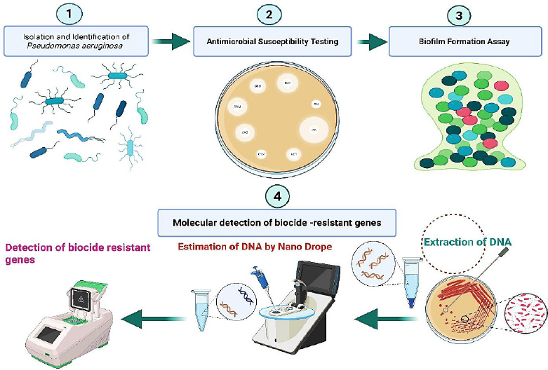
A diagram describing the experimental procedures for isolation, antibiotic susceptibility testing, biofilm measurement, and genetic profiling of qac genes in P. aeruginosa. This figure was created by BioRender.
2.2. Bacterial Isolation and Identification
A total of 50 non-duplicate clinical isolates of P. aeruginosa were collected from various clinical sources at the laboratories of Ramadi Teaching Hospital from June 2025 to December 2025. Routine diagnostic specimens were used to recover clinical isolates (urine, blood, wound, burn) of P. aeruginosa and cultured onto MacConkey agar and cetrimide agar (HiMedia Laboratories Pvt. Ltd., Mumbai, India, Cat# M081, M024). Incubation of plates was done at 37°C. The initial identification was done using morphological characters of the colonies, pigment generation, and oxidase test. Final confirmation was performed using the automated Vitek-2 apparatus as per the instructions of the manufacturer [15].
2.3. Antimicrobial Susceptibility Testing
Antibiotic susceptibility profile was assessed using the Kirby-Bauer disk diffusion test on Mueller-Hinton agar medium (HiMedia Laboratories Pvt. Ltd., Mumbai, India, Cat# M173) according to CLSI recommendations. Standardization of bacteria was done to 0.5 McFarland and spread onto agar plates. Aseptically, antibiotic disks were used, and plates were incubated at 37°C for 18 -24 h. Measures were made of inhibition zones, and susceptibility was interpreted according to CLSI breakpoints. Many antibiotic discs were used in this test (HiMedia, India), including: ciprofloxacin (CIP), ceftazidime (CAZ), piperacillin (PIP), piperacillin/ tazobactam (TZP), gentamicin (GEN), cefepime (FEP), amikacin (AMK), imipenem (IPM), and colistin (COL) (HiMedia Laboratories Pvt. Ltd., Mumbai, India). The classification of MDR and XDR was according to international standards [16, 17].
2.4. Biofilm Formation Assay by 1% Crystal Violet Method
Biofilm formation was assessed in triplicate utilizing standard microtiter plates (Corning Inc., Corning, NY, USA, Cat# 3596). The overnight cultures were diluted 1: 100 in tryptic soy broth (HiMedia Laboratories Pvt. Ltd., Mumbai, India, Cat# M011) and inoculated in sterile 96-well plates (200 µL/well). Following incubation at 37°C (24 h), wells were washed with PBS (HiMedia Laboratories Pvt. Ltd., Mumbai, India, Cat# TL1101), then they were allowed to air-dry and fixed with methanol. The biofilms were fixed with 0.1% crystal violet (HiMedia Laboratories Pvt. Ltd., Mumbai, India, Cat# GRM961) for 20 min, rinsed with deionized water, and destained with 95% ethanol (RCI Labscan Ltd., Bangkok, Thailand, Cat# UN1170). Measurement of absorbance was done at 570 nm. Isolates were classified as weak, moderate, and strong producers [18].
2.5. Antiseptic Tolerance Testing
Broth microdilution was used in the determination of tolerance to chlorhexidine (HiMedia Laboratories Pvt. Ltd., Mumbai, India, Cat# RM1254) and benzalkonium chloride (HiMedia Laboratories Pvt. Ltd., Mumbai, India, Cat# GRM751). Serial dilutions were done twice in a microtiter plate then, the bacterial suspensions were inoculated to 5x10⁵ CFU/ml. Plates were incubated at a temperature of 37 °C. The growth of bacteria was observed visually, and the minimum bactericidal concentration (MBC) was noted. The MBC was determined by the visual end-point in which no bacterial growth was recovered on subsequent subcultures. Isolates were assigned to discrete tolerance categories based on fixed MBC thresholds: ‘Susceptible’ (complete growth inhibition at standard working dilutions: ≤0.02% v/v for chlorhexidine, and ≤0.01% w/v for benzalkonium chloride); ‘Reduced Tolerance’ (bacterial survival up to 2-fold above standard concentrations); and ‘High Tolerance’ (bacterial persistence at concentrations ≥4-fold above standard clinical working dilutions) [19].
2.6. Molecular Study
2.6.1. Extraction of Genomic Bacterial DNA
Genomic DNA extraction was performed on fifteen biocide-resistant P. aeruginosa isolates using the bacterial DNA extraction kit (Geneaid Biotech Ltd., New Taipei City, Taiwan, Cat# GBB100 / GBB101), adhering to the provided instructions. Fifteen isolates were selected based on their highest levels of antibiotic resistance, strongest biofilm-forming capacity, and enhanced tolerance to biocides and antiseptic agents.
2.6.2. Quantitative Estimation of DNA Concentration and Purity by UV-Nanodrop Spectrophotometer Technique
Following the extraction of the genomic DNA, the concentration and purity of each of the DNA samples were determined by the NanoDrop spectrophotometer. To calibrate the instrument, 1 µL of TE buffer (HiMedia Laboratories Pvt. Ltd., Mumbai, India, Cat# ML016) was used to blank. Then 1 µL of genomic DNA of each isolate was placed on the measurement pedestal and measured based on instructions given by the manufacturer (Thermo Fisher Scientific, USA).
2.6.3. Selection and Design of Primers for Biocides Resistant P. aeruginosa Genes
The biocide resistance genes qacE and qacEΔ1 were detected through polymerase chain reaction (PCR), employing previously established primer sequences derived from the study Kazama et al.: for qacE (365bp), forward primer 5'-ATG AAA GGC TGG CTT-3' and reverse primer 5'-TTA GTG GGC ACT TGCTTT GG-3; for qacEΔ1(300 bp), forward primer 5'-TAG CGA GGG CTT TAC TAA GC-3' and reverse primer 5'-ATT CGA AAT GCC GAA CAC CG-3' (Bioneer, Daejeon, South Korea) [20].
2.6.4. Preparation of Primers
The primers used in this study were supplied by Bioneer Company in a lyophilized state, with varying picomolar concentrations. To prepare them for use in experiments, the primers were reconstituted using nuclease-free water (TransGen Biotech, Beijing, China, Cat# GBB100 / GBB101), resulting in a final concentration of 10 picomoles per microliter of solution.
2.6.5. PCR Detection of qacE and qacEΔ1 Genes
The polymerase chain reaction (PCR) amplification was performed utilizing DNA amplification kit (TransGen Biotech, Beijing, China, Cat# AS111-01) to detect qacE (365 bp) and qacEΔ1 (300 bp) genes using previously described primers. Each reaction contained template DNA, primers, dNTPs, MgCl2, Taq polymerase, and buffer in a final volume of 25 µL. PCR cycle conditions were as follows: initial denaturation at (95°C for 5 min.), 35 cycles of denaturation at (95 °C for 30 sec.), annealing at (55 °C for 30 sec.), and extension at 72 °C for 1 min.), followed by a final extension at (72 °C for 5 min.). Finally, PCR products were quantified on 1.5 agarose gel (HiMedia Laboratories Pvt. Ltd., Mumbai, India, Cat# MB002) stained with ethidium bromide (HiMedia Laboratories Pvt. Ltd., Mumbai, India, Cat# MB071) [20].
2.7. Biosafety and Chemical Safety
The University of Anbar and Ramadi Teaching Hospital's institutional biosafety and chemical safety regulations were followed in all routine laboratory operations, biocide handling, and disposal procedures.
2.8. Statistical Analysis
Data analysis of this study was performed using GraphPad Prism software (version 8). The Chi-square test was used to assess the relationships among antibiotic resistance, biofilm strength, antiseptic tolerance, and gene carriage. Associations between variables were assessed using Odds Ratio (OR) with 95% Confidence Intervals (CI) to determine effect strengths. Logistic regression models were employed to investigate whether patterns of multidrug resistance predicted higher tolerance range to antiseptics. Statistical significance was set at p < 0.05.
3. RESULTS
3.1. Antibiotic Resistance Panel among Surveyed Pseudomonas aeruginosa
Antibiotic testing results showed that the most pronounced resistance was towards fluoroquinolones and a number of β-lactam agents, whereas colistin and carbapenem activity remained mainly conserved (Fig. 2). Ciprofloxacin exhibited the poorest antimicrobial activity overall: 39 isolates (78.0%) were resistant, only 7 isolates (14.0%) were sensitive, and 4 isolates (8.0%) showed moderate to low sensitivity. Ceftazidime displayed high resistance, with 32 isolates (64.0%) resistant, 5 (10.0%) intermediate, and 13 (26.0%) susceptible. More than half of the isolates demonstrated a similar pattern to piperacillin, with 26 (52.0%) being resistant, 4 (8.0%) intermediate, and 20 (40.0%) susceptible.
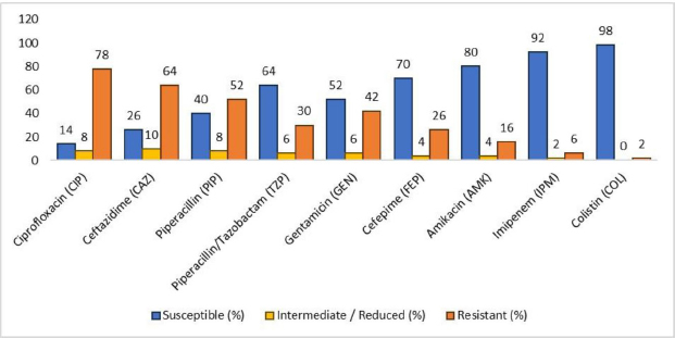
Relative distribution of antibiotic susceptibility among P. aeruginosa isolated (n=50). Susceptibility testing was evaluated according to CLSI criteria. Clear differences were observed between highly resistant classes (fluoroquinolones and β-lactam) and highly resistant agents (carbapenems and polymyxins).
In contrast, higher susceptibility rates were recorded for aminoglycosides and β-lactam combinations. Piperacillin/tazobactam showed sensitive activity in 32 isolates (64.0%) and a low resistance rate (15; 30.0%). Cefepime retained good activity, with 35 isolates (70.0%) susceptible and 13 (26.0%) resistant. Gentamicin showed a mixed pattern: 21 (42.0%) were resistant, 3 (6.0%) were intermediate, and 26 (52.0%) isolates were sensitive.
Amikacin, imipenem, and colistin were found to be the most effective. The susceptibility of amikacin was 80.0% (40 isolates), with 16.0% (8 isolates) were resistance. Imipenem remained highly effective, as there were 46 (92.0%) isolates being susceptible and only 3 (6.0%) being resistant. Colistin was almost active, with 49 isolates (98.0%) sensitive and one isolate (2.0%) resistant, meaning that it can still be used as a last-line choice in this set of isolates.
3.2. Classification of Bacterial Resistance based on Antibiotics Panel
Pseudomonas aeruginosa isolates showed an evident prevalence of multidrug-resistant strains (Table 1). Among 50 isolates that underwent analysis, 32 (64.0%) were identified as MDR, meaning that they were resistant to more than three antimicrobial classes, which indicates a high antimicrobial resistance burden among the clinical isolates. Non-MDR isolates accounted for 15 cases (30.0%), which is less than a third of the entire collection and indicates there is a low ratio of strains without altered antimicrobial susceptibility. The XDR isolates were present at a low rate, and only 3 isolates (6.0%) were found, but the occurrence is clinically important because of the critical limitation of treatment that is linked to this phenotype. Altogether, the results demonstrate a high rate of MDR among P. aeruginosa isolates, reflecting the persistence problem of antimicrobial resistance in clinical practice.
| MDR Status | Number of Isolates | Percentage (%) |
|---|---|---|
| Non-MDR | 15 | 30.0% |
| MDR | 32 | 64.0% |
| XDR | 3 | 6.0% |
| Total | 50 | 100% |
3.3. Classification of Bacterial Isolated based on their Biofilm Formation
The biofilm formation analysis of the isolates of P. aeruginosa indicated that most of them formed biofilms to varying extents (Table 2). Strong biofilm development seen in 19 isolates (38.0%), denoting a significant ability for surface adhesion and persistence. Fifteen isolates (30.0%) exhibited moderate biofilm production, and the same number of isolates displayed weak biofilm production. Only one isolate (2.0%) showed no detectable biofilm formation. These results indicated that the ability of biofilm-forming was common among the studied isolates, which may contribute to boosted resistance and survival in clinical environments.
| Biofilm Category | Number of Isolates | Percentage (%) |
|---|---|---|
| None | 1 | 2.0% |
| Weak | 15 | 30.0% |
| Moderate | 15 | 30.0% |
| Strong | 19 | 38.0% |
| Total | 50 | 100% |
3.4. Antiseptic Tolerance Profile in P. aeruginosa Isolates
P. aeruginosa isolates showed a variable response to the tested biocidal agents (Table 3). Susceptibility to antiseptics was observed in 20 isolates (40.0%), representing the largest single group. A decrease in tolerance was observed in 15 isolates (30.0%), indicating some adaptation to antiseptics exposure. The proportion of isolates with high-level tolerance was 15 (30.0%) (high CHX (n=5), high BAC (n=4), high all (n=6)), indicating that a significant fraction of the isolates has the capacity to resist antiseptics in common use. All of these results indicate that 60% of the isolates possessed some extent of reduced susceptibility, which is indicative of the possible difficulty of antiseptic tolerance in clinical practice.
| Antiseptic Response | Number of Isolates | Percentage (%) | ||
|---|---|---|---|---|
| Susceptible | 20 | 40.0% | ||
| Reduced tolerance | 15 | 30.0% | ||
| High tolerance | High CHX | 5 | 15 | 30.0% |
| High BAC | 4 | |||
| High all | 6 | |||
| Total | 50 | 100% | ||
3.5. Correlation between Antimicrobial Resistance Phenotypes and Antiseptic Tolerance Patterns in P. aeruginosa
There was a strong correlation between the status of antimicrobial resistance and antiseptic tolerance (Table 4). Susceptible and reduced susceptibility were observed in all non-MDR isolates. On the contrary, MDR isolates were spread across all types of tolerance, such as reduced susceptibility and high-tolerance phenotype. It is important to note, however, that all XDR isolates were highly tolerant to all the introduced antiseptics. The results of the Chi-square test proved that there is a strong relationship between status of resistance and antiseptic tolerance (χ2= 63.47, df = 8, p < 0.0001), which means that the higher the antimicrobial resistance, the greater the tolerance to CHX, BAC, and combined antiseptics.
| MDR Status | Susceptible | Reduced | High CHX | High BAC | High All | Total | |
|---|---|---|---|---|---|---|---|
| Non-MDR | 11 | 4 | 0 | 0 | 0 | 15 | |
| MDR | 9 | 11 | 6 | 5 | 1 | 32 | |
| XDR | 0 | 0 | 0 | 0 | 3 | 3 | |
| Total | 20 | 15 | 6 | 5 | 4 | 50 | |
| χ2, df | 63.47, 8 | ||||||
| p-value | <0.0001 | ||||||
| p-value summary | **** | ||||||
3.6. Correlation between Biofilm Formation Strength and Antimicrobial Resistance Status (in P. aeruginosa Isolates)
A statistically significant association was observed between antimicrobial resistance status and biofilm-forming capacity (Fig. 3). Non-MDR isolates predominantly exhibited weak biofilm formation (14/15), with only one isolate demonstrating moderate biofilm production and none classified as strong biofilm producers. Conversely, the shift of MDR isolates was towards increased biofilm formation, and most of them formed moderate (13/32) to strong (17/32) biofilms, while only two MDR isolates exhibited weak biofilm formation. On the same note, the XDR isolates (n = 3) had a greater tendency towards biofilm formation since two of the three isolates formed strong biofilms, and the remaining one had moderate biofilm formation. Generally, the high levels of biofilm formation were mostly linked to MDR and XDR phenotypes, whereas the low levels of biofilm formation were predominantly linked to non-MDR isolates.
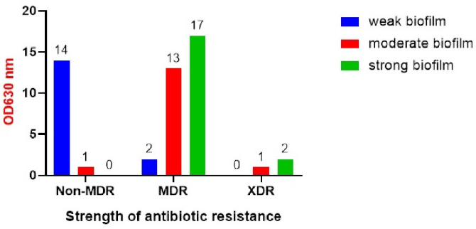
Relationship between antimicrobial resistance strains and biofilm formation strength. Comprehensive analysis shows high-level biofilm patterns (moderate to strong) are specifically associated with MDR and XDR strains, while weak biofilm production clusters within non-MDR isolates. The statistical significance was determined using chi-square analysis (χ2 =37.44, df = 4, p < 0.0001).
Overall, the strongest biofilm formation was the most common phenotype (n = 19), whilst the weak (n = 16) and the moderate (n = 15) biofilm formers were less frequently observed. The analysis of the statistical data showed that the status of antimicrobial resistance and the strength of biofilm formation have a significant association (χ2 = 37.44, df = 4, p < 0.0001), which implies that high antimicrobial resistance in P. aeruginosa is closely related to high biofilm-forming ability.
3.7. Correlation between Biofilm Formation and Antiseptic Tolerance
It was evident that there was a relationship between biofilm-forming capacity and resistance to antiseptics (Table 5). Non-forming isolates of biofilm and weak biofilm formers were virtually all vulnerable, with no apparent tolerance to chlorhexidine (CHX), benzalkonium chloride (BAC), or a combination of the antiseptics. Conversely, moderate and high biofilm producers had an increasing degree of antiseptic tolerance. In moderate biofilm formers, the majority of the isolates displayed susceptibility or decreased susceptibility, with a minor percentage displaying high tolerance to all the antiseptics. The most resistant were strong biofilm formers, with some of the isolates being very tolerant to CHX, BAC, or all the antiseptics used. The chi-square test as a statistical test established that there was a very significant correlation between biofilm strength and antiseptic tolerance (χ2= 53.05, df =12, p < 0.0001).
| Biofilm | Susceptible | Reduced | High CHX | High BAC | High All | Total |
|---|---|---|---|---|---|---|
| None | 1 | 0 | 0 | 0 | 0 | 1 |
| Weak | 14 | 1 | 0 | 0 | 0 | 15 |
| Moderate | 5 | 9 | 0 | 0 | 1 | 15 |
| Strong | 0 | 5 | 5 | 4 | 5 | 19 |
| Total | 20 | 15 | 5 | 4 | 6 | 50 |
| χ2, df | 53.05, 12 | |||||
| p-value | <0.0001 | |||||
| p-value summary | **** | |||||
3.8. Molecular Detection of Biocide Genes among P. aeruginosa Isolates
Following the extraction of DNA, the concentration of genomic DNA ranged from 70-300 ng/uL, and A260/ A280 purity ratio was found to be 1.80-1.98, as measured using a NanoDrop spectrophotometer. Fifteen clinical isolates of P. aeruginosa were measured about their high antibiotic resistance levels, high biofilm-forming ability, and significant resilience to antiseptic agents.
All fifteen (100%) P. aeruginosa isolates screened in this study carried the qacEΔ1 gene, as confirmed by amplification of the specific 300 bp PCR product (Fig. 4).
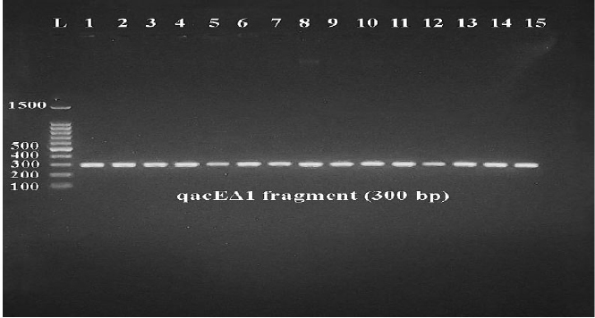
Agarose gel electrophoreses for amplified qacEΔ1 fragment (300bp). M: 100 bp DNA ladder; L1-L8: PCR products of P. aeruginosa DNA using P. aeruginosa primers.
Among the fifteen P. aeruginosa isolates analyzed, eleven (73.3%) were positive for the qacE gene, yielding the expected amplicon size of 365 bp, whereas four isolates (26.7%) showed no detectable amplification of this gene (Fig. 5). These results indicated that the presence of qacE was strongly associated with a high antiseptic resistance profile (11 positive vs 4 negative isolates; p = 0.011, odds ratio = 12.5 (95% CI:1.34-116.42)), suggesting an apparent direct contribution to biocidal survival (Table 6).
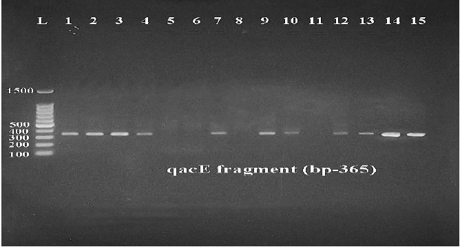
Agarose gel electrophoresis for amplified qacE fragment (365bp). M: 100 bp DNA ladder; L1-L12: PCR products of P. aeruginosa DNA using P. aeruginosa primers.
| Isolate ID | MDR/XDR Status | Biofilm Category | Antiseptic Tolerance Profile | qacE | qacEΔ1 |
|---|---|---|---|---|---|
| Iso 1,2,3 | XDR | Strong | High tolerance (CHX/BAC) | Positive | Positive |
| Iso 4,7,9,10,12,13,14,15 | MDR | Strong/ Moderate | High tolerance (CHX/BAC) | Positive | Positive |
| Iso 5,6,8,11 | MDR | Moderate | Reduced tolerance | Negative | Positive |
| Odds Ratio | 12.5 (95% CI:1.34-116.42) | ||||
| p-value | <0.011 | ||||
| p-value summary | **** | ||||
4. DISCUSSION
This study presents a comprehensive phenotypic and genotypic characterization of the determinants of antiseptic resistance in clinical isolates of P. aeruginosa. It has shown a worrying convergence of strong biofilm formation ability, extensive drug and antiseptic resistance, along with a significant prevalence of qac genes. The substantially higher incidence of qacEΔ1 (100%) compared to qacE (73.3%) aligns with previous studies demonstrating that qacEΔ1 is widely distributed among Gram-negative bacteria due to its persistent integration within a class 1 integron [21, 22]. QacEΔ1, while regarded as a shortened derivative of qacE, maintains functional activity and imparts diminished susceptibility to quaternary ammonium compounds, chlorhexidine, and benzalkonium chloride [23]. The significant statistical correlation between qacE and qacEΔ1 carriage and biocide resistance (p < 0.01) emphasizes the notion that extended selective pressure from regular hospital antiseptic application fosters the proliferation of gene-bearing strains. Furthermore, the frequent co-occurrence of qac genes with antibiotic resistance determinants within class 1 integrons offered a genetic explanation for the emergence of MDR and XDR strains [24]. In this investigation, qacEΔ1 was primarily found in MDR and XDR isolates, which is consistent with global epidemiological patterns that link mobile genetic platforms to efflux-mediated biocide resistance. This phenomenon poses a significant challenge to infection control, because tolerance to antiseptics may indirectly contribute to the survival and development of antibiotic-resistant strains even in the absence of direct antimicrobial exposure [25].
In this study, most isolates (98%) exhibited biofilm-forming capacity. This is consistent with the well-known ability of P. aeruginosa bacteria to form biofilms [26]. It is known that biofilm-associated communities promote horizontal gene transfer and provide up to a 1,000-fold enhancement in antimicrobial tolerance [27]. The highly statistically significant association between biofilm strength and multidrug resistance/extreme drug resistance status (p < 0.0001) indicated that biofilm formation is another defense mechanism that increases the persistence of antibiotic efficacy. It's interesting to observe that isolates bearing qacEΔ were overrepresented as potent biofilm producers, as qac-mediated efflux capacity has been observed to enhance the resistance of sessile cells to antiseptics [28]. The efflux mechanisms play a dual role in biocidal resistance and maintaining cellular homeostasis, promoting biofilm growth and survival of P. aeruginosa bacteria on hospital surfaces, indwelling catheters, water systems, and medical equipment [29].
Concurrently, both MDR and XDR isolates exhibit remarkable tolerance to popular antiseptics such as sodium hypochlorite, benzalkonium chloride, chlorhexidine, and ethanol. The clinical importance of biocide-induced selection pressure was highlighted by the fact that XDR isolates in particular were consistently found to be extremely tolerant to all antiseptics in use [30]. According to the results of this study, the strong biofilm producer strains were more likely to have high levels of tolerance to CHX, BAC, or a combination of them (p < 0.0001). These findings align with mechanistic evidence that suggests qac-encoded efflux pumps forcibly expel biocidal chemicals from the bacterial cell while the extracellular polymeric matrix guarantees that antiseptic penetration is limited. The synergistic action of these mechanisms creates a multilayered defense system that enables P. aeruginosa to survive standard hospital disinfection practices [31, 32].
From an epidemiological and infection control perspective, the ability to form biofilm and widespread biocide resistance due to the high prevalence of qacE and qacEΔ1 indicates the shortcomings of the current disinfection technique in healthcare facility environments [33]. The risk of transmissibility of the class 1 integrons harboring qac genes is especially significant in high dependency units like intensive care units and burn wards, where intense selective pressure creates appropriate conditions for microbial survival and outbreak progression [34, 35]. The results highlight the need to combat the impact of antiseptic tolerance and antibiotic resistance using combined stewardship programs, and the interaction between co-selection and the emergence of multidrug resistance [36, 37]. Despite the strength of the molecular and phenotypic data presented in the current study on the biocide resistance mechanisms, additional studies should be undertaken to clarify the genetic background and evolutionary processes of biocide-antibiotic co-resistance in P. aeruginosa through sequencing the integron structures, analysis of efflux pump expression, and whole-genome sequencing.
5. LIMITATIONS
Despite P. aeruginosa being a highly adaptable and dangerous pathogen in hospitals, this study has a few key limitations. Further investigations into gene expression levels using real-time q-PCR are needed to determine the activity of resistance genes and the relationship between these levels and antibiotic resistance. Expanded studies (in vivo) using laboratory animals are recommended because they are closer to human tissues than laboratory environments. The molecular screening of qacE and qacEΔ1 was performed on a subset of 15 highly tolerant isolates, rather than the complete 50-isolate collection. While this improved our understanding of resistance determinant co-selection, it introduced a selection bias and prevented a realistic baseline estimate of absolute qac gene prevalence across all isolated strains. Expanding thorough genotypic screens to include all separated specimens is a priority for future studies. Incorporating advanced microscopy techniques remains a vital direction for future structural studies to visually validate phenotypic tolerance profiles.
CONCLUSION
This study indicates the presence of a strong connection between antiseptic resistance determinants, biofilm formation, and multi-drug resistance in P. aeruginosa. The prevalence of qacE and qacEΔ1, especially among MDR and XDR strains, is very high and highlights the importance of integron-associated genes in the decreased susceptibility to the widely used antiseptics. The ability to form biofilms was closely associated with antimicrobial resistance and antiseptic tolerance, with organisms producing robust biofilms showing the greatest level of resistance. The MDR and XDR isolates were found to have much higher tolerance to chlorhexidine, benzalkonium chloride, and combined antiseptics, implying that resistance in P. aeruginosa is preconditioned by the synergistic action of biofilm-mediated resistance and efflux-related mechanisms. These results imply that daily disinfection measures can also promote the continued spread of drug-resistant strains. Thus, the proper use of antiseptics and close monitoring of biocide resistance markers should be incorporated within infection control programs, which also involves antibiotic stewardship.
AUTHORS’ CONTRIBUTIONS
The authors confirm their contribution to the paper as follows: S.A.A. and B.H.K.: Proposed the presented idea; S.A.A.: Created the hypothesis and performed the calculations; N.Q.J.: Confirmed the analysis approaches; S.A.A. B.H.K., and N.Q.J.: Experimented and contributed to the final version of the manuscript. All authors reviewed the results and approved the final version of the manuscript.
LIST OF ABBREVIATIONS
| CLSI | = Clinical and Laboratory Standards Institute |
| qac gene | = quaternary ammonium compounds gene |
| MDR | = Multi-Drug Resistant |
| XDR | = Extensively Drug-Resistant |
| PCR | = Polymerase Chain Reaction |
| PBS | = Phosphate-Buffered Saline |
| CFU/mL | = Colony Forming Unit per Milliliter |
| MBC | = Minimum Bactericidal Concentration |
| DNA | = Deoxyribonucleic Acid |
| TE | = Buffer Tris-EDTA Buffer |
| CHX | = Chlorhexidine |
| BAC | = Benzalkonium Chloride |
| dNTPs | = Deoxynucleoside Triphosphates |
ETHICS APPROVAL AND CONSENT TO PARTICIPATE
The protocol was reviewed and approved by the Institutional Ethics Committee of the University of Anbar, Iraq (Approval Reference No. 221 on November 23, 2025).