All published articles of this journal are available on ScienceDirect.

Association between Microbial Colonization and Spontaneous Abortion: A Clinical Study of Post-Curettage Patients
 iD
Osamah Faisal Kokaz2
iD
Athraa A. Abdulrab4
iD
iD
Osamah Faisal Kokaz2
iD
Athraa A. Abdulrab4
iD
Authors Info & Affiliations
Abstract
Introduction
Spontaneous abortion causes approximately 50,000 maternal deaths annually, primarily due to microbial infection and hemorrhage, accounting for 8–13% of global maternal deaths. This study aimed to investigate the association between microbial infections, bacterial diversity, and patterns related to spontaneous abortion in two groups of women.
Methods
A total of 156 bacterial isolates were recovered from 48 patient samples, while 67 isolates were obtained from 48 healthy controls. All samples were cultured under aerobic and anaerobic conditions on enrichment and selective media, including Blood agar, MacConkey agar, and Chocolate agar.
Results
Gram-positive bacteria predominated among total isolates (53.84%) compared to Gram-negative bacteria (46.15%). The primary Gram-positive pathogens were S. aureus (29.16%), L. acidophilus (27.08%), Peptostreptococcus spp. (22.91%), and C. perfringens 20.83%. Among Gram-negative isolates, E. coli was most prevalent (47.91%), followed by P. aeruginosa (18.75%) and K. pneumonia (16.6%). In the control group, L. acidophilus (43.75%) and E. coli (18.75%) were most common. Diversity analyses showed no significant difference in α-diversity, but β-diversity revealed clear distinctions between groups. Linear Discriminant Analysis identified specific biomarkers differentiating the microbial compositions of the two cohorts.
Discussion
These findings indicate a Gram-positive predominance, with Staphylococcus aureus and E. coli identified as the primary pathogenic drivers in the study groups. Although species richness remained consistent across cohorts, β- diversity and Linear Discriminant Analysis biomarkers confirmed distinct microbial shifts and bacterial signatures between the examined groups.
Conclusion
L. acidophilus appears to help maintain vaginal health and reduce the risk of spontaneous abortion, as β- diversity and biomarker analyses revealed a shift from beneficial to pathogenic bacteria in women with spontaneous abortion.
1. INTRODUCTION
Miscarriage and premature delivery represent significant challenges in women’s reproductive health, with significant implications for maternal and fetal outcomes. Miscarriage is defined as the spontaneous termination of a pregnancy prior to 24 weeks of gestation. It is typically categorized into early miscarriage (embryonic loss before 12 weeks) and late miscarriage (fetal loss between 12 and 24 weeks) [1-3]. According to the World Health Organization (WHO), however, spontaneous miscarriage can be classified into four clinical stages: threatened, inevitable, incomplete, and complete [4].
Reported evidence suggests that alterations in the vaginal microbiota may play a critical role in adverse pregnancy outcomes. Conditions such as Bacterial Vaginosis (BV), various Sexually Transmitted Diseases (STDs), preterm birth, gynecological malignancies, premature pre-labor membrane rupture, Polycystic Ovary Syndrome (PCOS), and recurrent implantation failure have all been associated with dysbiosis of the vaginal microflora [5-7]. Recent studies have highlighted an association between reduced microbial diversity and richness in the vaginal microbiota and an increased risk of miscarriage [8] and preterm birth [9, 10]. Notably, a decline in Lactobacillus spp., together with a corresponding rise in microbial diversity, has been linked to spontaneous preterm delivery [11, 12].
The vaginal microbiota is an important factor in regulating immunity throughout the female reproductive tract [13]. Alterations in the vaginal microbiota may cause immunological dysregulations, potentially leading to an increased susceptibility to adverse pregnancy outcomes, including miscarriage. For instance, infection of human endometrial cells by Chlamydia trachomatis has been associated with defective decidualization and reduced chemokine production, both of which may contribute to miscarriage [14, 15].
Genital tract infections are also a well-documented cause of pregnancy loss. According to Bjartling et al. [16], infections associated with abortions frequently result from an increased rate of bacterial infection, including chlamydia, gonorrhea, mycoplasma, and BV. Such infections typically originate in the lower genital tract and progress via the cervix to reach the uterus; if left untreated, they can disseminate to the fallopian tubes and result in infertility. Several infections have been correlated with an elevated risk of miscarriage. The literature indicates that women who have experienced a miscarriage are more commonly affected by systemic infections, including syphilis, brucellosis, and HIV, as well as viral and parasitic infections such as cytomegalovirus, dengue fever, rubella, and malaria [1].
In terms of normal flora, a variety of bacteria, including Diphtheroid, Staphylococcus epidermidis, streptococci, and Escherichia, constitute the vaginal microbiota during the prepubertal years [17]. Vaginal colonization by Group B Streptococcus, which triggers epithelial exfoliation and the subsequent upward migration of bacteria, is linked to a higher incidence of spontaneous miscarriage [18]. A recent study linked pregnancy loss to a significant reduction in vaginal Lactobacillus spp. abundance during the first trimester [19]. Another study found that unexplained recurrent miscarriage was associated with higher abundances of Atopobium, Streptococcus, and Prevotella, whereas healthy controls showed higher abundance of Lactobacillus and Gardnerella [20].
These pathogens can cause severe infections following unsafe abortion, which leads to the hospitalization of 7 million women worldwide [21]. Hemorrhage, infection, retained fetal products, amniotic fluid embolism, vaginal tract injury, uterine perforation, and cervical laceration are among the most serious complications that can arise from unsafe abortions [22, 23]. Furthermore, several studies have closely linked Spontaneous Abortion (SA) and abnormal vaginal flora and associated vaginitis, likely through inflammatory mechanisms affecting pregnancy outcome [24]. The exact mechanisms by which BV might cause SA remain unclear; diminished chorioamniotic function may result from changes in host defenses that permit ascending intrauterine infection, which in turn damages chorioamnion formation [25]. Similarly, embryo damage or SA may occur following cytokine activation, which increases prostaglandin production, negatively affecting the endometrium and fetal growth [26].
It has been hypothesized that Group B Streptococcus, a common colonizer of the maternal reproductive tract, contributes to adverse outcomes by secreting virulence factors that weaken gestational tissues [27]. Group A Streptococcus infection, although infrequent, is linked to a swift and severe progression of puerperal sepsis and a rapidly advancing soft tissue infection [28]. The overall mortality rate among patients with Group A Streptococcus infection is approximately 3%, rising to 4% in the presence of septic shock [29, 30].
This study aims to investigate the relationship between microbial infections, bacterial diversity, and the pattern associated with spontaneous abortion in two groups of women.
2. MATERIALS AND METHODS
2.1. Sample Collection
A total of 96 vaginal swabs were collected, comprising 48 from the spontaneous abortion (patient) group and 48 from a healthy control group in a balanced 1:1 design. All participants were admitted to Shomaly Teaching Hospital, South of Babylon, Iraq, between January and December 2022. Samples were collected at the time of miscarriage while patients were hospitalized. All patients in the study group had experienced at least one miscarriage. The age range was 18-37 years for the patient group, and 18-38 years for the healthy control group. The control group consisted of pregnant women with regular menstrual cycles prior to pregnancy and moderate sexual activity.
2.2. Inclusion and Exclusion Criteria
Women were eligible for inclusion in the patient group if they were of reproductive age (18–37 years), female, and had experienced spontaneous abortion. Women were eligible for inclusion in the control group if they were of reproductive age (18–38 years), pregnant with a normal physiological status, had regular menstrual cycles prior to pregnancy, and reported moderate sexual activity. Women with serious underlying diseases were excluded from participation, as were those who declined to provide consent.
Vaginal swabs were collected following standard guidelines, and verbal informed consent was obtained from all participants prior to sample collection. The study was approved by the Iraqi Ministry of Health. Samples were obtained by a gynecologist in the examination room, with participants in the lithotomy position, immediately following miscarriage. To collect an adequate quantity of vaginal fluid, synthetic swabs were rotated three times within the posterior vaginal fornix. The specimens were then immediately transferred and stabilized for 10 minutes, then frozen at -20°C for further identification.
A post-hoc power analysis was conducted using G*Power 3.1. With an alpha level of 0.05 and a sample size of 48 participants per group, the study achieved sufficient statistical power (0.80) to detect a medium-to-large effect size of (d = 0.58) between the patient and control groups.
2.3. Microbial Culture and Identification
Vaginal swabs were inoculated onto enrichment and selective media under both aerobic and anaerobic conditions. Samples were cultured on blood agar, MacConkey agar, and chocolate agar to isolate pure colonies, followed by Gram staining for morphological characterization. To differentiate between bacterial species, biochemical assays, including catalase, oxidase, urease, and coagulase tests, were performed on suspicious colonies [31]. Final species identification was confirmed using the VITEK-2 diagnostic system (bioMérieux, France) [32].
2.4. Statistical Analysis
Statistical data analysis was performed using IBM SPSS. Significant differences in α-Diversity were assessed using the Mann-Whitney U test, and β- diversity was evaluated using three metrics: Jaccard Distance, Bray-Curtis Dissimilarity, and Sørensen-Dice Distance. Additionally, Linear Discriminant Analysis Effect Size (LEfSe) was applied, with statistical significance set at P < 0.05. Python was used to perform Principal Coordinates Analysis (PCoA) of Jaccard distances for aborted and healthy cohorts.
3. RESULTS
In this study, the bacterial communities within the vaginal ecosystem served as the primary basis for comparison between the patient group and the healthy control group. A total of 156 isolates were obtained from 48 swab samples collected from the patient group, identified based on cultural characteristics and biochemical testing. In contrast, 67 isolates were accurately identified from the 48 samples of the healthy control group. Within the patient group, Gram-positive bacteria were dominant, comprising 84 isolates (53.84%), while Gram-negative bacteria accounted for 72 isolates (46.15%). The healthy control group yielded 67 bacterial isolates, of which 41 (61,19%) were Gram-positive, and 26 (38.8%) were Gram-negative.
Among the patient group (ages 18–37), the predominant Gram-positive isolates were Staphylococcus aureus (n = 14, 29.16%), Lactobacillus acidophilus (n = 3, 27.08%), and Peptostreptococcus spp. (n = 1, 22.91%), as shown in Fig. (1). Other species identified in this group included Clostridium perfringens (n = 10, 20.83%), Staphylococcus epidermidis (n = 7, 14.58%), and Group A and B Streptococci (n = 6, 12.5%). Less prevalent species included Enterococcus faecalis (Group D Streptococcus) (n = 2, 4.16%) and Bifidobacterium spp. (n = 5, 10.41%), Listeria monocytogenes (n = 3, 6.25%), Corynebacterium spp. (n = 3, 6.25%), and Clostridium botulinum and Ureaplasma urealyticum (n = 2, 4.16%).
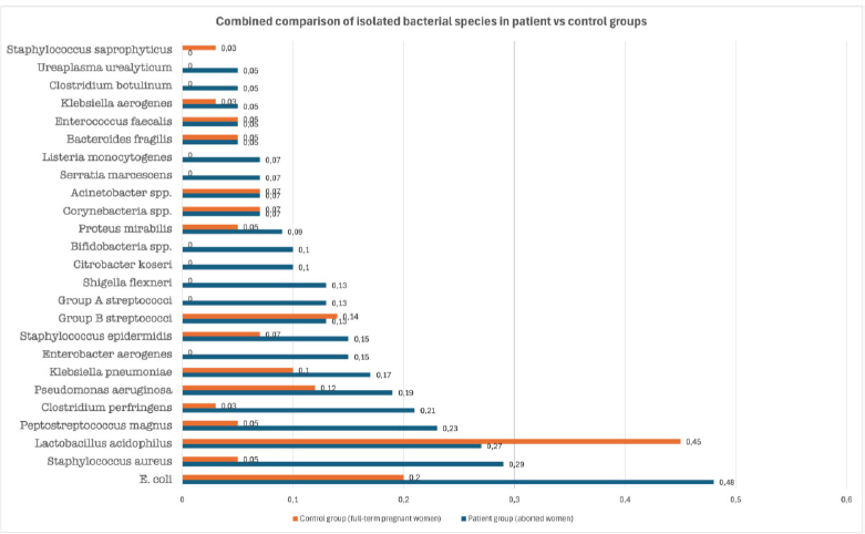
Comparative distribution of bacterial species isolated from patient and control groups. Horizontal bars represent the approximate proportion of each bacterial species isolated from the patient group (women with spontaneous abortion) and the control group (women with full-term pregnancies). Orange bars represent the patient group and the blue bars represent the control group. Species not detected in a given group were assigned a value of zero to enable direct visual comparison across all organisms. Data are plotted in descending order of frequency within the patient group.
Gram-negative bacteria (Fig. 1) also maintained a significant presence in the microbial ecosystems of aborted women across different age groups. Escherichia coli was the most frequently isolated species at (n = 23, 47.91%), followed by Pseudomonas aeruginosa (n = 9, 18.75%), Klebsiella pneumoniae (n = 8, 16.6%), Enterobacter aerogenes (n = 7, 14.8%), Shigella flexneri (n = 6, 12.5%), Citrobacter koseri (n = 5, 10.41%), Proteus mirabilis (n = 4, 8.3%), Actinobacteria spp., and Serratia marcescens (n = 3, 6.25%). In rare cases, Bacteroides fragilis and Klebsiella aerogenes were found in 2 isolates each (4.16%).
The reference group (Fig. 1) consisted of 48 healthy women (ages 18–38) admitted to Shomaly Teaching Hospital during the same period as the case group. Laboratory and biochemical testing identified 67 isolates; Gram-positive species accounted for 41 (61.19%), while Gram-negative species accounted for 26 (38.8%). The vaginal ecosystem in these women was rich in bacteria associated with hormonal and physiological status. Seven genera formed the Gram-positive components, with the highest frequencies being L. acidophilus (n = 21, 43.75%) and Group B streptococci (n = 6, 12.5%), consisting of S. agalactiae (6.25%), S. pyogenes (4.16%), and S. pneumoniae (2%). Other isolates included Corynebacterium spp. and S. epidermidis (n = 3, 6.25% each), Staphylococcus aureus, Peptostreptococcus magnus, and Enterococcus faecalis (n = 2, 4.16% each), and Clostridium perfringens and Staphylococcus saprophyticus (n = 1, 2% each).
Gram-negative bacteria were also interspersed within the normal flora of the vaginal mucosa. Seven genera were identified: E. coli was predominant (n = 9, 18.75%), followed by Pseudomonas aeruginosa (n = 5, 10.41%), Klebsiella pneumoniae (n = 4, 8.3%), Actinobacteria (n = 3, 6.25%), Proteus mirabilis and Bacteroides fragilis (n = 2, 4.16% each), and Klebsiella aerogenes (n = 1, 2%).
Alpha-diversity testing indicated no significant difference in richness between the case and healthy groups as assessed by the Mann-Whitney U test (P = 0.3943, above the 0.05 threshold).
Beta-diversity revealed significant differences in microbial structure between aborted and healthy women. The case group displayed a more variable and complex composition, characterized by dysbiosis of beneficial bacteria. Specifically, the presence of Clostridium perfringens (OR= 12,37, 95% CI [1.51, 100.99]), Staphylococcus aureus (OR = 9,47, 95% CI [2.02, 44.43]), and Peptostreptococcus spp. (OR = 6.84, 95% CI [1.43, 32,82]) showed strong positive associations with the case group. Conversely, Lactobacillus acidophilus was inversely associated with the case group (OR = 0.48, 95% CI [0.20, 1.13]), reflecting its prevalence within the normal vaginal flora of the healthy reference group. According to Jaccard distance and Bray-Curtis dissimilarity indices (P < 0.05), the case group displayed a more variable and complex composition due to the dysbiosis of beneficial bacteria such as Lactobacillus spp. This dysbiosis appeared to favor the growth of pathogenic and opportunistic species, including Staphylococcus aureus, Peptostreptococcus spp., and Clostridium perfringens. Conversely, the Sørensen-Dice distance showed low dissimilarity, indicating that overall species similarity remained high between the two study groups, as shown in Table 1. Patho-microbial changes may be influenced by several factors, including the timing of abortion (early or late pregnancy); furthermore, these pathogens may potentially disseminate via the bloodstream to other critical sites.
Table 1.
| Test | Value | Range | Description | Explanation |
|---|---|---|---|---|
| Jaccard Distance | 0.4 | 0-1 | Moderate dissimilarity (60% similarity) | Presence/Absence patterns |
| Bray-Curtis dissimilarity | 0.546 | 0-1 | Substantial dissimilarity (40% similarity) | Abundance weighted composition |
| Sørensen-Dice Distance | 0.25 | 0-1 | Low-moderate dissimilarity (75% similarity) | Shared species emphasis |
The Jaccard distance (Fig. 2) demonstrates the distribution of aborted women (A) and healthy women (H) in a three-dimensional space. The blue clusters, representing samples from the case group, are concentrated in the center, while the orange clusters, representing healthy women, are located around the periphery with relatively lower density. This provides a clear advantage for observing abundance in the Bray-Curtis dissimilarity test, even though the number of detected genera remained non-significant, as detailed in the Sørensen-Dice distance test.
Linear Discriminant Analysis Effect Size (LEfSe), with an LDA threshold score > 4.0, was performed to identify biomarkers distinguishing the case and control groups. Aborted women reached the threshold score for three genera: Peptostreptococcus, Clostridium perfringens, and Listeria monocytogenes. Other species with lower scores associated with vaginal dysbiosis and pregnancy complications included S. aureus, E. aerogenes, Group A streptococci, and Ureaplasma urealyticum. In comparison, healthy women showed significantly higher levels of Lactobacillus acidophilus than the case group, suggesting a protective role against opportunistic bacteria, alongside Group B streptococci and P. aeruginosa, which are components of the normal vaginal flora at moderate abundance, as summarized in Table 2.
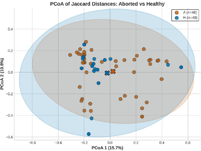
Principal coordinates analysis (PCoA) of jaccard distances for aborted and healthy cohorts.
Table 2.
| Bacteria | LDA Score (effect size) |
Abundance in Aborted Group |
Bacteria | LDA Score (effect size) |
Abundance in Healthy Group |
|---|---|---|---|---|---|
| *P. meganus | 4.31 | Higher | *L. acidophilus | 4.65 | Higher |
| *C. perfringens | 4.22 | Higher | *Group B streptococci | 4.41 | Higher |
| *S. aureus | 3.98 | Higher | *P. aeuroginosa | 4.02 | Higher |
| E. aerogenes | 3.87 | Higher | K. pneumonia | 3.84 | Higher |
| L. monocytogenes | 3.52 | Higher | Corynebacteria | 3.56 | Higher |
| Group A streptococci | 3.48 | Higher | Actinobacteria | 3.44 | Higher |
| U. urealyticum | 3.15 | Higher | B. fragilis | 3.23 | Higher |
| K. aerogenes | 2.89 | Higher | P. mirabilis | 2.97 | Higher |
| S. marcescens | 2.75 | Higher | S. epidermidis | 2.63 | Higher |
| C. koseri | 2.54 | Higher | - | - | - |
| S. flexneri | 2.33 | Higher | - | - | - |
| C. botulinum | 2.11 | Higher | - | - | - |
4. DISCUSSION
To date, numerous studies have established that the vaginal microbiota maintains a close relationship with spontaneous abortion and preterm delivery. An imbalance or decrease in Lactobacillus prevalence, observed at 27.08% in women who experienced abortions compared to 47.75% in healthy women, plays a direct role in pregnancy loss, particularly during the first trimester, consistent with findings from [19]. This shift can weaken the protective physiological role of Lactobacillus spp. within the reproductive tract [33]. Furthermore, vaginal dysbiosis is associated with an increase in proinflammatory cytokines [34], as the reduction in Lactobacillus populations facilitates the overgrowth of harmful pathogens [35]. The link between Lactobacillus disruption and premature delivery was previously investigated by Elovitz et al. [5], confirming that women with normal pregnancies typically possess a stable microbiota dominated by Lactobacillus [36].
Many aerobic bacteria residing in the reproductive mucosa, such as Corynebacterium, Escherichia, Enterococcus, and Streptococcus (all isolated in this study), are associated with aerobic vaginitis and urinary tract infections. These organisms negatively affect women undergoing in vitro fertilization. Furthermore, pathogenic bacteria such as E. faecalis, E. coli, S. agalactiae, and Gardnerella vaginalis proliferate in the absence of adequate Lactobacillus richness [37]. A study by Fu et al. [6] on women with unexplained recurrent implantation failure demonstrated that Lactobacillus abundance is positively associated with pregnancy rates and the resolution of vaginal dysbiosis.
Salmanov et al. [38] isolated various bacteria from women experiencing abortion, vaginitis, and endometritis in Ukraine. The most frequent isolate was E. coli (25.9%), consistent with the current study's findings, which identified it as the leading Gram-negative isolate at 47.91%. Other isolates included Enterococcus (16.2%), S. aureus (15.5%), P. aeruginosa (10.9%), E. aerogenes (10.1%), Actinobacteria (4.9%), and P. mirabilis (2.5%); these findings closely mirror those observed in the aborted women of the present study. These microorganisms also show high prevalence in vaginal samples from patients with urinary tract infections [38].
The vaginal flora is less complex than the intestinal flora; in women of childbearing age, it comprises approximately 40 species of aerobic and anaerobic bacteria, including Lactobacillus, Bacteroides, Corynebacterium, and Escherichia [39]. Statistical analysis in this study demonstrated a significant association (p < 0.05) between Bacterial Vaginosis (BV) and a history of spontaneous abortion. Notably, 70.6% of women diagnosed with BV had experienced at least one spontaneous abortion [40]. This finding aligns with recent evidence reporting a diverse range of bacterial species isolated from women with a history of abortion. An imbalanced vaginal microbiota, characterized by reduced Lactobacillus and anaerobic dominance, may contribute to preterm delivery and low birth weight.
DNA sequencing and data analysis of miscarriage samples demonstrated an overrepresentation of Lactobacillus and Gardnerella in control groups compared with women experiencing recurrent miscarriages. This suggests that an abundance of taxa such as Atopobium, Streptococcus, and Prevotella is directly linked to bacterial vaginosis and subsequent adverse birth outcomes [20]. Additionally, the abundance of Group B streptococci in the case group of aborted women suggests a role in intrauterine infection and spontaneous abortion. These findings emphasize that microbial imbalance fundamentally results from pathogenic and/or opportunistic bacteria proliferating beyond normal levels within the vaginal community [1].
Technological developments have advanced our understanding of the microbiome; however, “omics” metrics, such as alpha- and beta-diversity measures related to disease, are not yet widely utilized by physicians [38]. Researchers have noted that different species within the Lactobacillus genus have dissimilar functions; specifically, L. crispatus and L. iners play distinct roles in community stability [39]. The current study found that alpha-diversity levels involving Lactobacillus spp. and other taxa showed no significant differences between groups; this may be influenced by hormonal regulation, though further investigation is needed to confirm this relationship. Furthermore, the decrease in Lactobacillus and increase in Prevotella, Clostridium, Bacteroides, and Dialister observed via LEfSe analysis of euploid miscarriage samples compared with aneuploid samples [15] are consistent with the current results. Further research is necessary to fully explore the specific roles of the normal vaginal flora in spontaneous abortion and miscarriage.
5. STUDY LIMITATIONS
This study is culture-based but lacks molecular analyses, which could have provided deeper insights into the underlying mechanisms. While we recognize that NGS data provide superior depth and precision, the absence of sequencing platforms at our institution posed a significant logistical barrier. Consequently, conducting such advanced genomic analyses was not feasible for this study. We have ensured that the current methodology is robust and that its limitations are transparently discussed.
CONCLUSION
The current study underscores the pivotal role of Lactobacillus acidophilus in maintaining and improving the vaginal microbiome to reduce the incidence of spontaneous abortion. Our findings reveal that L. acidophilus abundance was significantly higher in the healthy control group compared with the aborted cohort, suggesting a protective effect.
Analysis of β-diversity and (LEfSe) Linear Discriminant Analysis Effect Size biomarkers further highlighted a marked shift in community composition; non-healthy (aborted) women exhibited a substantial increase in pathogenic-genus diversity, whereas healthy women were characterized by a dominance of beneficial bacteria. These results suggest that depletion of Lactobacillus species may precede the proliferation of opportunistic pathogens, ultimately compromising pregnancy stability.
AUTHORS’ CONTRIBUTIONS
The authors confirm their contribution to the paper as follows: D.A.M., H.A.: Study conception and design; D.A.M., A.A.A., O.F.K.: Data collection; D.A.M., H.A.: Analysis and interpretation of results; D.A.M., H.A., A.A.A., O.F.K.: Draft manuscript. All authors reviewed the results and approved the final version of the manuscript.
LIST OF ABBREVIATIONS
| BV | = Bacterial Vaginosis |
| STDs | = Sexually Transmitted Diseases |
| PCOS | = Polycystic Ovary Syndrome |
| WHO | = World Health Organization |
| SA | = Spontaneous Abortion |
| IVF | = In Vitro Fertilization |
| OR | = Odds Ratio |
| CI | = Confidence Interval |
| LDA | = Linear Discriminant Analysis |
| LEfSe | = Linear Discriminant Analysis Effect Size |
| NGS | = Next-Generation Sequencing |
| SPSS | = Statistical Package for the Social Sciences |
ETHICS APPROVAL AND CONSENT TO PARTICIPATE
The research protocol was approved by the Ethical Committee of Al-Shomaly hospital belongs to the Babylon Health Directorate.
This study was also approved by the Ethics Committee of the Iraqi Ministry of Health, in compliance with all national regulations. The project approval number and date were 74,590, issued on December 1, 2022.
HUMAN AND ANIMAL RIGHTS
All procedures involving human participants were conducted in accordance with the ethical standards of the committee responsible for human experimentation (institutional and national), and with the Helsinki Declaration of 1975, as revised in 2013.
CONSENT FOR PUBLICATION
Verbal informed consent was obtained from all participants prior to sample collection.
AVAILABILITY OF DATA AND MATERIALS
All data generated or analyzed during this study are included in this published article.
ACKNOWLEDGEMENTS
The authors would like to thank Shomaly Teaching Hospital, South of Babylon, Iraq, for their support and cooperation during the sample collecting process.