All published articles of this journal are available on ScienceDirect.

Colchicine Induced Mutation in Nigella sativa Plant for the Assessment of Morpho-Physiological and Biochemical Parameter Vis-A-Vis In Vitro Anti-Inflammatory Activity
Authors Info & Affiliations
Abstract
Background:
Nigella sativa (NS), an herbaceous medicinal plant recognized for its diverse beneficial applications as a spice and traditional medicine.
Objective:
The present study was targeted to explore the antioxidant potential of Nigella sativa in response to colchicine-induced mutation. The stress condition brought due to mutation may affect the medicinal value (anti-inflammatory activity) of the plant.
Method:
Nigella sativa seeds were imperiled to colchicine treatment at various concentrations viz. 0.00625, 0.0125, 0.025, 0.05 and 0.1% subjected for analysis.
Result:
The colchicine treated plant (polyploid/ mutant) at 0.025% concentrations showed significant variation at morpho-physiological and biochemical level with respect to control (p value < 0.05). At the morphological level, the plant showed enlargement of shoot length (33.760±2.295mm), root length (13.546±1.535 mm), and leaf area (22.836±1.524 mm2). The analysis of seeds showed enhanced seeds per pod (49.333±4.163), weight of seeds (2.810±0.010g), length (3.133±0.089mm), and width (1.123±0.044mm) when compared with control. The physiological parameters also showed significant enhancement for stomatal index (35.456±4.751%), chlorophyll A (9.053±0.865 µg/gfw), chlorophyll B (4.990±0.763 µg/gfw), and total carotene content (773.190±5.906 µg/gfw). However, the fresh weight/ dry weight ratio (10.738±3.031) was found to be deprived. Furthermore, biochemical parameters viz. total flavonoid (seeds 1.973±0.134; plant 1.703 ± 0.064 mg eqv QE/g of tissue), total phenolic (seeds 15.690±1.495; plant 8.220±0.070 mg eqv GA/g of tissue), total carotene (seeds nil; plant 773.190±5.906 µg/gfw), and total antioxidant (seeds 0.445±0.102; plant 0.386±0.010 mM eqv AA/g tissue) were significantly elevated at 0.025% of colchicine treatment. When the in vitro anti-inflammatory activity was targeted, a significant escalation was observed for inhibition of albumin denaturation (97.466±2.835%), proteinase inhibitory activity (62.290±6.475%), heat-induced hemolysis (89.873±3.533%), hypotonicity induced hemolysis (92.572±3.527%), anti-lipoxygenase activity (96.010±3.098%), and cyclooxygenase inhibitory activity (68.296±3.920%) at 500µg/mL concentration of extract.
Conclusion:
Thus, it can be concluded that 0.025% of colchicine can induce significant (p value < 0.05) mutation in the Nigella sativa plant, which may lead to alterations at morpho-physiological and biochemical levels. Such treatment induces stress in the plant and leads to elevated antioxidant levels. This in turn elevates the therapeutic potential of the plant. Hence, our study is a novel and open-ended finding to explore various other medical properties of the plant with respect to colchicine-induced mutation.
1. INTRODUCTION
The therapeutic plant, Nigella sativa belonging to the Ranunculaceae family, is a profound miracle herb with a rich historical and religious background. Extensive research investigations had previously revealed its wide spectrum pharmacological potential [1, 2]. Nigella sativa is commonly known as the black seed, which is a native plant to Southern Europe, North Africa, and Southwest Asia, including India. Nigella sativa is an annual flowering plant that grows to 20–30 cm (7.9–11.8 inches) tall and has linear-lanceolate leaves. The delicate flowers have 5-10 petals and the colors are usually yellow, white, pink, pale blue, or pale purple. The fruit of the plant is a large and inflated capsule composed of 3-7 united follicles with numerous seeds. The black-colored seeds are flattened, oblong, angular, and funnel-shaped with a length of 0.2 cm and 0.1 cm wide [3]. Most of the beneficial properties of this plant is because of the presence of secondary metabolites (namely thymoquinone, dithymoquinone, thymohydroquinone, and thymol) having antioxidant potential [4]. The whole plant is beneficial for health, but its seeds and its oil are considered to be most important, as they possess active phytochemical constituents [5]. This plant has an immense therapeutic potential, which has been used for curing diseases for many centuries in different native systems of medicine [2].
Nigella sativa seeds and its oil showed significant anti-inflammatory activity, which is primarily attributed to the therapeutic compound thymoquinone [6]. Nigella sativa seed extracts are also shown to have pro-apoptotic and anti-inflammatory effects in pancreatic cancer cells [7]. Nigellone is another phytochemical of Nigella sativa which shows the inhibitory action on the release of histamine from mast cells; thus, exhibiting anti-asthmatic property [8]. According to previous observations, the extract of the Nigella sativa plant showed anti-inflammatory activity in animal systems. In the study, the alcoholic extract of Nigella sativa seeds and callus obtained from the leaves of the plant was used to determine anti-inflammatory activity [9]. It was shown to have an anti-inflammatory effect on mix glial cells of rats with respect to its thymoquinone content at different concentrations. The study showed that thymoquinone content in the callus of the leaf has 12 times higher anti-inflammatory activity than alcoholic extract of seeds. It was observed in similar studies that the dose of thymoquinone has a time-dependent effect. Furthermore, it significantly decreases tumor necrosis factor-alpha, the pancreatic ductal adenocarcinoma cell synthesis of monocyte chemoattractant protein-1, cyclooxygenase, and interleukin (IL)-1 β [9].
Less work has been done in the propagation of therapeutic plants through breeding techniques. Here a biotechnological technique like polyploidization may trigger up breeding and development of novel mutants. Among other techniques, polyploidization is mostly practiced in asexually propagated plants to evolve a variety with some different characters like leaf size, plant size, bud size, stomata count [10]. In the last few decades, polyploids have been successfully produced in many ornamental crops [11]. Polyploidy breeding proves to be a very efficient technique in comparison to conventional cross-breeding and mutation breeding. Ease in handling and short span of time increasing the germplasm availability render polyploidy breeding its advantage over conventional methods [12, 13]. Furthermore, the mutation caused by artificial polyploidization involves gene mutation, which results in whole-genome modification, thus, creating more phenotypic variations associated with single gene mutation [14]. Moreover, it is found that polyploidy does not occur logically in all genera of plants, so it is induced artificially in most of the economically significant crops [15]. Induced polyploids provide an effective method to improve genetic variation in the area of plant breeding and genetics [16]. Most research works showed that colchicine induced polyploidy acts interspecifically, resulting in hybridization or somatic cell doubling [17]. Mitotic polyploidization includes the doubling of chromosomes in somatic cells. This chromosome doubling is broadly used to encourage tetraploids using anti-mitotic chemicals [18]. Though, synthetically produced sexual polyploids are often used for breeding determination.
Since 1930, colchicine is used as a mutagen in the plant breeding technique, that can induce mutation /polyploidy in plants [19]. The mechanism of action of colchicine involves the inhibition of microtubules formation and doubling the chromosome number. It is widely used to make polyploid plants and works as a mitotic poison by introducing mutagenic effects [20]. Colchicine induces polyploidy by inhibiting the chromosome segregation throughout meiosis, which gives half of the gametes (sex cells) thereby, altering the chromosome number. It is clearly stated that colchicine not only promotes the alteration of chromosome number but also induces mutation in plants. Colchicine treatment affects the phytochemical production in plants, thereby affecting the medicinal property of the plant. With consideration to the medicinal property of Nigella sativa and the effect of mitotic inhibitor (colchicine) inducing polyploidy/ mutation, the present investigation was designed. The investigation would evaluate the effect of induced mutation on phytochemicals synthesis of plant and thus affecting the medicinal value (anti-inflammatory activity) of the plant. The mutant plants having an enhanced level of phytochemicals of therapeutic importance could be beneficial for the pharmaceutical and nutraceutical industry for herbal drug formulations.
2. MATERIALS AND METHODS
2.1. Plant Material and Colchicine Treatment
Reference seeds were collected from the National Research Centre on Seed Spices Tabiji Rajasthan, India. The seeds were grown at the Botanical garden of Amity University Uttar Pradesh, Lucknow Campus. Control and colchicine treated seeds were sown in late October and harvested in early March. The various morphological, physiological, and biochemical studies were conducted on seeds and plants of Nigella sativa. Colchicine treatments were given to Nigella sativa seeds at a concentration of 0.00625, 0.0125, 0.025, 0.05 and 0.1% for 8 hours each. 100 seeds of Nigella sativa were taken and soaked in a petri plate lined with filter paper. For control experiments, the seeds were soaked in distilled water. After treatment with colchicine, the seeds were transferred to the Botanical garden of Amity University Uttar Pradesh, Lucknow campus for germination and development of plants for further analysis. Primarily, morphological analysis was performed to determine whether colchicine-induced mutation was effective or not. Finally, the physiological, cytological, and biochemical study was performed.
2.2. Morphological Study
The collected seeds were used to study the variation in morphological features, especially the size of seeds, the weight of seeds, and seeds per pod. For whole-plant analysis, the parameters such as root length, shoot length, and leaf area were estimated using the protocol of Iqbal et al. [21]. Morphological features were examined under compound binocular microscope Optika Digital microscope and Magnus MLX microscope and photographed using a digital camera. Readings were estimated in triplicates and the mean± standard deviation was calculated.
2.3. Physiological Study
Physiological parameters were calculated according to the protocol of Iqbal et al. [21]. The plants obtained after 30 days of germination were studied for traits like average root and shoot length and average leaf area. The fresh weight and dry weight ratio of the seedlings were calculated by taking the average weight of 10 random plants. Dry weight was taken after incubating the plants at 72°C for 18 hours. Chlorophyll A, chlorophyll B, and total carotene content were determined spectrophotometrically with some modifications [22]. The weighed samples were homogenized in 100% acetone and the homogenate was filtered through Whatman No.1 filter paper and was further centrifuged using the REMI cooling centrifuge C-24BL model at 2500 rpm for 10 min. The supernatant was collected and the absorbance was taken at 662 nm (for chlorophyll A), 646 nm (for chlorophyll B), and 470 nm (for total carotene) using Shimadzu UV- 1800 spectrophotometer. The calculations were performed according to the formulae of Lichtentaler and Wellburn [23] as mentioned below.
Chlorophyll a = 11.75 A662 - 2.350 A645
Cholorophyll b = 18.61 A645 - 3.960 A662
Total carotene = 1000 A470 - 2.270 Ca - 81.4 Cb/227
Ca = Chlorophyll a, Cb = Chlorophyll b
Readings were taken in triplicates and the mean ± standard deviation was calculated. The values were denoted as μg/g fresh weight (μg/gfw).
Further, the stomatal index was calculated according to the standard protocol of Paul et al. [24]. The formula used for the calculation of the stomatal index is as follows:
Stomatal index % = (Number of stomata/Number of stomata + Number of epidermal cell) X 100
2.4. Cytological Study
For the cytological study, the protocol of Tsuchiya and Nakamura [25] was used with minor modifications. Root tips were pretreated with ice-cold water overnight. The sample material was fixed in a fixative solution containing ethanol and glacial acetic acid in a ratio of 3:1 for 24 hours. For the mitotic study, the root tips were collected from a fixative solution and transferred on a watch glass for hydrolytic maceration using several drops of 1N HC1 at 60°C for 6 min. Thereafter, 1N HCl was removed using blotting paper, and the sample was washed with distilled water for the removal of extra 1N HCl. Staining was done by dipping the root tips in 0.8 to 1.0% acetocarmine solution for 15 min. After incubation tip was covered with a coverslip, and then blotting paper was placed over the coverslip and pressed gently with a thumb to squash the plant material. The slide was heated for few seconds over the flame. The chromosomes were studied under microscopic observation using a binocular microscope (OPTIKA microscope Italy, B-383Phi) and finally photographed.
2.5. Biochemical Study
2.5.1. Total Flavonoids
Total flavonoid was extracted by the method of Iqbal et al. [26]. 1 gram of tissue was submerged in 4 mL of 80% ethanol overnight. The extract was filtered twice using Whatman no. 1 filter paper. The filtrate was collected in fresh tubes and placed into storage at –20 °C until further analysis. Ethanolic extracts were taken and kept at room temperature, vortexed, and diluted to 1:10 with 80% ethanol. Absorbance was taken at 362 nm using a Shimadzu UV- 1800 spectrophotometer. Readings were taken in triplicates and the mean (± standard deviation) was calculated to plot the graph. Flavonoid quantification was done by using Quercetin (QE) as standard.
2.5.2. Total Phenolic
Total phenolic content was determined by the protocol of Iqbal et al. [26], with some modifications. Tissue weight (1 gram) was extracted by homogenizing with 10 mL of 70% ethanol. The extract was filtered twice with Whatman filter paper No 1. The collected filtrates were pooled and evaporated to dryness and finally stored at 4 °C until use. Gallic acid was used as a reference standard for plotting the calibration curve. A volume of 0.5 mL of the plant extract was mixed with 2 mL of the Folin-Ciocalteu reagent (diluted 1:10 with de-ionized water) and was neutralized with 4 mL of sodium carbonate solution (7.5%, w/v). The reaction mixture was incubated at room temperature for 30 min with intermittent shaking for color development. The absorbance of the resulting color was measured at 765 nm using a UV- VIS spectrophotometer in triplicate. The total phenolic content was determined from the linear equation of a standard curve prepared with gallic acid (GA). Readings were taken in triplicates and the mean (± standard deviation) was calculated.
2.5.4. Total Antioxidant Activity
Total antioxidant activity was assayed by the protocol of Cacig et al. [27], with some modifications. 1 gram of tissue was homogenized in 4 mL of double-distilled water and incubated for 24 hours at 4°C. It was then filtered twice with Whatman No. 1 filter paper and the filtrate collected was stored at 4°C. A suitable amount of sample was taken in a 3mL glass cuvette containing the oxidative mixture of 0.18 mL potassium permanganate (0.01M), 0.42 mL sulfuric acid (2M), and 2.4 mL distilled water. The decrease in absorbance was measured at 535 nm. Ascorbic acid (AA) was taken as standard. Readings were taken in triplicates and the mean activity (± standard deviation) was calculated. To quantitatively compare the antioxidant activities, formula of Iqbal et al. [28] was used.
2.6. Therapeutic Study (In vitro Anti-inflammatory Activity)
2.6.1. Inhibition of Protein (Albumin) Denaturation
To quantify the protein (albumin) denaturation, the protocol of Sakat et al. [29] and Rastogi et al. [30] was used with minor alterations. The study was performed with an equivalent volume of 1% bovine albumin (Fraction V) and 0.025% colchicine treated plant extracts. The pH was adjusted using 1 N HCl. At 37°C, incubation was done for 20 min followed by 51°C temperature for 20 min to terminate the reaction. Later, the reaction mixture was cooled at RT and optical density was taken at 660 nm using ultraviolet (UV)-visible spectrophotometer (Shimadzu UV-1800). Aspirin was used as a reference drug. The calculation for percentage inhibitory activity of protein (albumin) denaturation using the following formula was applied.
% inhibition = [{Control Abs–Sample Abs}/Control Abs] × 100
2.6.2. Antiproteinase Activity
To measure the activity of antiproteinase, the protocols of Oyedepo and Femurewa [31] and Sakat et al. [29], were followed with some modifications. In 0.025% colchicine treated plant extracts, 1 mM pH 7.4 Tris-HCl buffer (1 mL), and 0.001% trypsin (2 mL) were added. This was followed by incubation for 5 min at 37°C. After incubation 0.02% (w/v) casein (1 mL) was added and incubated for 20 min. Finally, 2% perchloric acid (2 mL) was added to stop the reaction. Thereafter, optical density was measured at 210 nm. As a reference drug, Aspirin was used and anti-proteinase activity was calculated using the following formula.
% inhibition = [{Control Abs.–Sample Abs}/Control Abs.] × 100
2.6.3. Heat-Induced Haemolysis
Heat-induced hemolysis activity was performed by adding an equal volume of plant extract (1 mL) and 10% RBCs suspension (1 mL). It was then incubated at 56°C in a water bath for 30 min. Thereafter, the reaction mixture was cooled at room temperature to stop the reaction and centrifuged (2500 rpm for 5 min) to get the supernatant. The absorbance was taken with supernatant at 560 nm. As a reference, aspirin drug was used. The percentage of heat-induced haemolysis was calculated using the following formula [29, 32].
% inhibition = [{Control Abs–Sample Abs}/Control Abs] × 100
2.6.4. Hypotonicity-Induced Haemolysis
The activity of hypotonicity-induced haemolysis was conducted by adding hyposaline (2 mL), phosphate buffer pH 7.0 (1 mL), and 10% RBCs suspension (0.5 mL) in the sample extract. The mixture was then incubated for 30 min at 37°C and finally centrifuged at 3000 rpm to get the supernatant. The absorbance of the supernatant was taken at 560 nm. As a reference, diclofenac sodium drug was used. The percentage of hypotonicity-induced haemolysis was calculated with presumptuous control as 100% using the following formula [30].
% protection = 100-(OD sample/OD control) × 100
2.6.5. Anti-lipoxygenase (LOX) Activity
The anti-lipoxygenase activity was measured by using the protocol of Shinde et al. [32] with some alterations. Linoleic acid was used as a substrate and lipoxidase was used as an enzyme. The reaction mixture was prepared with 2M borate buffer of pH 9.0 and lipoxidase enzyme solution having a concentration of 20,000 U/mL (0.25 mL) with 0.025% colchicine treated Nigella sativa plant extract, which is kept for incubation at 25°C for 5 min. After incubation, 1.0 mL of 0.6 mM linoleic acid was added and vortexed to stop the reaction. As a reference, indomethacin drug was used. At 234nm, absorption was taken and activity was calculated using the following formula.
% inhibition = [{Control Abs–Sample Abs}/Control Abs] × 100
2.6.6. Cyclooxygenase Activity
The activity was performed by using the procedure of Viji and Helen [33] with minor alterations. The reaction mixture was prepared with Tris-HCl buffer, glutathione, hemoglobin, and enzyme. The reaction was initiated by the addition of arachidonic acid and then incubated at 37°C for 20 min. The reaction was completed by adding 0.2 mL of 10% tricarboxylic acid in 1 N HCl. Thereafter, 0.2 mL of tert-butyl alcohol was added, followed by incubation for 20 min in a boiling water bath. Finally, the mixture was centrifuged (1000 rpm/5 min) and the supernatant was collected. The absorbance was taken at 632 nm. As a reference, ibuprofen drug was used. The activity was calculated with the below formula.
% inhibition = [{Control Abs–Sample Abs}/Control Abs] x 100
Table 1.
| Samples | Analysis of Seeds | Analysis of Plants | |||||
|---|---|---|---|---|---|---|---|
| Colchicine Treatment |
Seeds per pod ± SD | Weight of 1000 seeds (gram) ± SD | Length of seeds (mm) ± SD | Width of seeds(mm) ± SD | Shoot length (cm) ± SD | Root length (cm) ± SD | Leaf area (mm2) ± SD |
| 0.00625% | 38.000 ± 2.000 | 2.226 ± 0.020 | 2.336 ± 0.215 | 1.113 ± 0.004 | 21.386 ± 1.900 | 5.326 ± 0.560 | 10.580 ± 1.150 |
| 0.0125% | 41.666 ± 2.516 | 2.540± 0.026 | 2.430 ± 0.164 | 1.113 ± 0.024 | 23.816 ± 2.202 | 10.200 ± 1.945 | 18.520 ± 1.611 |
| 0.025% | 49.333 ± 4.163 | 2.810± 0.010 | 3.133 ± 0.089 | 1.123 ± 0.044 | 33.760 ± 2.295 | 13.546 ± 1.535 | 22.836 ± 1.524 |
| 0.05% | 28.666 ± 0.577 | 2.126 ± 0.020 | 2.723 ± 0.025 | 1.116 ± 0.057 | 23.330 ± 2.556 | 6.000 ± 1.590 | 13.896 ± 1.911 |
| 0.1% | 23.666 ± 1.527 | 2.076 ± 0.058 | 2.076 ± 0.058 | 1.116 ± 0.089 | 13.633 ± 2.616 | 2.966 ± 1.311 | 9.470 ± 0.854 |
| Control | 40.666 ± 1.527 | 2.030± 0.030 | 2.693 ± 0.228 | 1.111 ± 0.038 | 23.276 ± 1.822 | 7.700 ± 1.225 | 15.803 ± 1.999 |
2.7. Statistical Analysis
All the data was denoted as mean ± standard deviation. One way or two way ANOVA was applied to test the level of significance at a p-value ≤ 0.05. Correlation analysis was also performed to determine a significant relationship between the various parameter studied using MS excel and GraphPad Prism software.
3. RESULTS
The present investigation showed significant results upon colchicine treatment of Nigella sativa seeds. The colchicine treatment causes various changes at the cellular, morphological, physiological, and biochemical levels. This probably affects the medicinal properties of Nigella sativa. Significant results were observed at morphological levels ( Table 1 and Fig. 1) of plants with maximum shoot length (33.760±2.295 cm) at 0.025% colchicine treatment. Contrastingly, compared to control (23.276±1.822 cm), the shoot length exhibited lesser growth. When the root length was compared, similar trend was observed with maximum root length (13.546±1.535 cm) at 0.025% of colchicine treated plant than control (7.700±1.225 cm). The leaf area also showed maximum results at 0.025% colchicine treatment with a value of 22.836 ± 1.524 mm2. While the control sample had a leaf area of 15.803±1.999 mm2. When post harvested seed analysis was done for morphological study, the seeds per pod were found maximum (49.333±4.163) at 0.025% colchicine treatment while control showed 40.666±1.527 seeds per pod. The average weight of 1000 seeds was found with maximum value (2.810±0.010 grams) at 0.025% colchicine treatment, while control was observed to have 2.030±0.030 grams. Seeds length and width were observed maximum at 0.025% colchicine treatment with 3.133±0.089 mm and 1.123±0.044 mm respectively. For control, seed length and width were found to be 2.693±0.228 mm and 1.111±0.038 mm respectively. When one-way ANOVA was applied for various morphological parameters studied, a significant difference was observed between various colchicine treatments with a p-value < 0.05. It thus elucidates the effect of different concentrations of colchicine treatment.
The physiological parameters also revealed significant results at 0.025% of colchicine treatment for chlorophyll A, chlorophyll B, and total carotene content with the maximum value of 9.053±0.086 µg/g fresh weight, 4.990±0.763 µg/g fresh weight, and 773.190±5.960 µg/g fresh weight respectively. Similarly, control showed chlorophyll A, chlorophyll B and, total carotene content to be 7.293±0.342 µg/g fresh weight, 3.696±0.519 µg/g fresh weight, and 683.533±10.035 µg/g fresh weight respectively. The stomatal index was again observed maximum at 0.025% colchicine treatment with a value of 35.456±4.751%, while control showed 27.736±3.421%. However, when fresh weight/dry weight ratio was observed, it was found to be maximum with the control sample (25.681±4.382) while 0.025% colchicine treatment plant sample showed relatively lesser values (10.738±3.031). The results are given in Table 2. When one-way ANOVA was applied for various physiological parameters, a significant difference between various colchicine treatments was observed with a p-value < 0.05. It thus elucidates the effect of different concentrations of colchicine treatment.
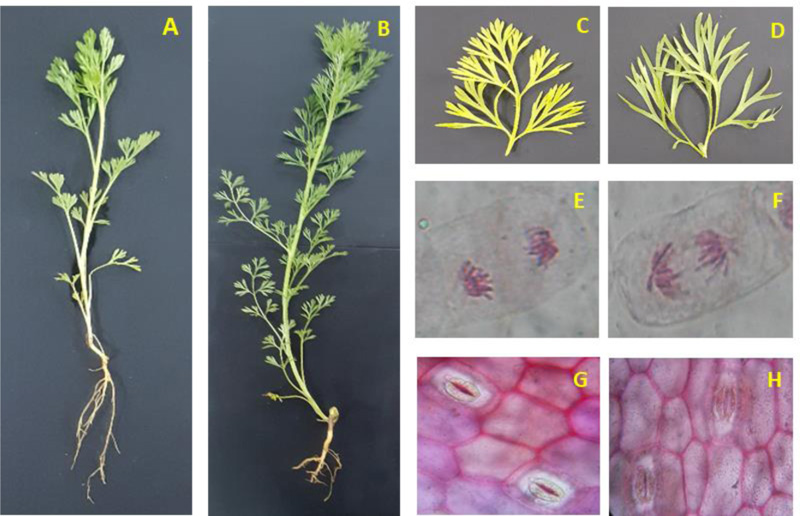
Table 2.
| Analysis of Plants | |||||
|---|---|---|---|---|---|
| Colchicine Treatment | Chlorophyll A Pigment µg /gfw | Chlorophyll B Pigment µg /gfw | Total Carotene (µg /gfw) | Stomatal Index % | Fresh Weight/Dry Weight ± SD |
| 0.00625% | 7.043 ± 0.715 | 4.530 ± 0.598 | 339.263 ± 6.780 | 25.176±3.902 | 21.578 ± 4.942 |
| 0.0125% | 8.676 ± 0.575 | 5.086 ± 0.152 | 648.366 ± 5.407 | 28.390±2.485 | 10.180 ± 0.836 |
| 0.025% | 9.053 ± 0.865 | 4.990 ± 0.763 | 773.190 ± 5.906 | 35.456±4.751 | 10.738± 3.031 |
| 0.05% | 8.280 ± 0.330 | 4.526 ± 0.188 | 629.843 ± 5.834 | 24.633±1.910 | 12.957 ± 9.812 |
| 0.1% | 8.243 ± 0.381 | 3.403 ± 0.393 | 468.280 ± 6.056 | 21.523±1.406 | 16.760 ± 1.713 |
| Control | 7.293 ± 0.342 | 3.696 ± 0.519 | 683.533 ± 10.035 | 27.736±3.421 | 25.681 ± 4.382 |
| Analysis of Harvested Seeds After Colchicine Treatment | Analysis of Plants | |||||||
|---|---|---|---|---|---|---|---|---|
| Colchicine Treatment | Total Flavanoids (mg eqv QE /g of Tissue) | Total Phenolics (mg eqv GA /g of Tissue) | Total Carotene (µg/gfw) | Total Antioxidant (mM eqv AA/g Tissue) | Total Flavanoids (mg eqv QE /g of Tissue) | Total Phenolics (mg eqv GA /g of Tissue) | Total Carotene (µg/gfw) | Total Antioxidant (mM eqv AA/g Tissue) |
| 0.00625% | 1.430 ± 0.160 | 9.173± 1.289 | _ | 0.380 ± 0.052 | 1.363 ± 0.040 | 5.380 ± 0.079 | 339.263 ± 6.780 | 0.205 ± 0.004 |
| 0.0125% | 1.633 ± 0.110 | 13.803 ± 1.335 | _ | 0.409 ± 0.115 | 1.470 ± 0.062 | 5.903 ± 0.107 | 648.366 ± 5.407 | 0.231 ± 0.009 |
| 0.025% | 1.973 ± 0.134 | 15.690 ± 1.495 | _ | 0.445 ± 0.102 | 1.703 ± 0.064 | 8.220 ± 0.070 | 773.190 ± 5.906 | 0.386 ± 0.010 |
| 0.05% | 1.066 ± 0.136 | 9.173 ± 0.619 | _ | 0.344 ±0.055 | 1.250 ± 0.060 | 5.823 ± 0.629 | 629.843 ± 5.834 | 0.190 ± 0.003 |
| 0.1% | 0.696 ± 0.126 | 6.306 ± 1.331 | _ | 0.209 ± 0.072 | 1.300 ± 0.200 | 4.426 ± 0.092 | 468.280 ± 6.056 | 0.110 ± 0.007 |
| Control | 1.800 ± 0.145 | 13.923 ± 0.640 | _ | 0.349 ± 0.044 | 1.463 ± 0.083 | 7.076 ± 0.051 | 683.533 ± 10.035 | 0.267 ± 0.051 |
The results showed significant variation at the cellular level as the chromosome density and stomatal structure varied in accordance with colchicine treatment. It can be observed from Fig. (1), the size and density of chromosomes have vividly increased after treatment with 0.025% colchicine when compared to the control sample. Further, the microscopic study elucidates the effect of colchicine treatment on the Nigella sativa plant. In the stomatal study, the number of stomatal count and epidermal cells showed significant variation between 0.025% colchicine treated plants and control plants sample. This might affect stomatal index values. The aforementioned characteristics of colchicine treated Nigella sativa plant led us to study its biochemical parameters. These parameters can be useful for phytochemical evaluation and therapeutic purposes.
The biochemical study for the Nigella sativa plant revealed significant results at 0.025% colchicine treatment for maximum total flavonoid, total phenolic, total carotene, and total antioxidant activity. The values recorded for the aforementioned parameters were 0.703±0.064 mg eqv QE/ g of tissue, 8.220±0.070 mg eqv GA/ g of tissue, 773.190±5.906 µg/ g fresh weight, and 0.386±0.010 mM eqv AA/ g of tissue, respectively. On the other hand, control sample for total flavonoid, total phenolic, total carotene, and total antioxidant activity showed deprived results with the value of 1.463±0.083 mg eqv QE/ g of tissue, 7.076±0.051 mg eqv GA/ g of tissue, 683.533±10.035 µg/g fresh weight, and 0.267±0.051 mM eqv AA/ g of tissue, respectively. The seeds obtained after harvesting the plant again showed significant results at 0.025% colchicine treatment for maximum total flavonoid, total phenolic, and total antioxidant activity. The values recorded for the aforementioned parameters were 1.973±0.134 mg eqv QE/ g of tissue, 15.690±1.495 mg eqv GA/ g of tissue, and 0.445±0.102 mM eqv AA/ g of tissue, respectively. For the control sample, the total flavonoid, total phenolic, and total antioxidant activity showed modest results with the value of 1.800±0.145 mg eqv QE/ g of tissue, 13.923±0.640 mg eqv GA/ g of tissue, and 0.349±0.044 mM eqv AA/ g of tissue, respectively. The results are given in Table 3. When one-way ANOVA was applied for various biochemical parameters, a significant difference between various colchicine treatments with a p-value < 0.05 was observed. It thus elucidates the effect of different concentrations of colchicine treatment.
The significant alterations due to colchicine treatment in Nigella sativa at various concentrations cause a stress response in the plant. However, it was observed that 0.025% colchicine treatment was most effective at morpho-physiological and biochemical changes. Therefore, the so-treated plant (0.025% colchicine treatment) was considered for its therapeutic (application) study. The in vitro anti-inflammatory activity was targeted with consideration to the antioxidant potential of the plant. The colchicine-treated plant samples showed better results than the control sample. It was observed that inhibition of albumin denaturation was maximum at 500µg/mL of extract used, with a value of 97.466±2.835% as compared to 87.500±3.541% in control. When proteinase inhibitory activity was measured, it was observed maximum at 500µg/mL of extract (62.290±6.475%) with respect to 43.790±5.970% in control. Heat-induced haemolysis revealed significant results with colchicine-treated extract (89.873±3.533%) at 500µg/mL of extract in comparison to control (68.006±3.208%). Hypotonicity-induced haemolysis showed higher activity (92.572±3.527%) than control (72.572±3.527%) at 500µg/mL of extract. The anti-lipoxygenase activity and cyclooxygenase inhibitory activity showed significant results with 96.010±3.098% and 68.296±3.920%, respectively. In contrast, the control samples showed anti-lipoxygenase activity and cyclooxygenase inhibitory activity of 88.610±4.061 and 79.530±4.664%, respectively, at 500µg/mL of extract. When the antioxidant potential was considered, it was found maximum with 85.577±3.049% in the treated sample and 77.100±3.256% in the control sample at 500µg/mL of extract. The results are given in Fig. (2). When two-way ANOVA was applied for various in vitro anti-inflammatory activity parameters, a significant difference was observed between various colchicine-treated samples and control samples with a p-value < 0.05. It thus elucidates the effect of colchicine treatment on the therapeutic property of the Nigella sativa plant.

| - | IAD S | PIA S | HIH S | HyIH S | LOX S | COX S | AA S | IAD C | PIA C | HIH C | HyIH C | LOX C | COX C | AA C |
|---|---|---|---|---|---|---|---|---|---|---|---|---|---|---|
| IAD S | 1 | |||||||||||||
| PIA S | 0.9282 | 1 | ||||||||||||
| HIH S | 0.9112 | 0.9202 | 1 | |||||||||||
| HyIH S | 0.9512 | 0.9965 | 0.9152 | 1 | ||||||||||
| LOX S | 0.9206 | 0.8727 | 0.7478 | 0.90793 | 1 | |||||||||
| COX S | 0.8796 | 0.9106 | 0.9862 | 0.90516 | 0.759 | 1 | ||||||||
| AA S | 0.9091 | 0.9761 | 0.946 | 0.97367 | 0.8649 | 0.9645 | 1 | |||||||
| IAD C | 0.9867 | 0.9613 | 0.9531 | 0.97483 | 0.90986 | 0.9399 | 0.963 | 1 | ||||||
| PIA C | 0.9523 | 0.9956 | 0.9086 | 0.99975 | 0.91069 | 0.8962 | 0.9684 | 0.973 | 1 | |||||
| HIH C | 0.9125 | 0.925 | 0.9999 | 0.91983 | 0.75374 | 0.9875 | 0.9506 | 0.9552 | 0.9133 | 1 | ||||
| HyIH C | 0.9512 | 0.9965 | 0.9152 | 1 | 0.90793 | 0.9052 | 0.9737 | 0.9748 | 0.9998 | 0.9198 | 1 | |||
| LOX C | 0.9512 | 0.9061 | 0.872 | 0.93295 | 0.96506 | 0.8913 | 0.937 | 0.9653 | 0.9305 | 0.876 | 0.933 | 1 | ||
| COX C | 0.9005 | 0.87 | 0.9821 | 0.87484 | 0.76238 | 0.9865 | 0.9289 | 0.9427 | 0.8664 | 0.9816 | 0.8748 | 0.9 | 1 | |
| AA C | 0.9709 | 0.9734 | 0.8986 | 0.98859 | 0.95538 | 0.8961 | 0.964 | 0.9844 | 0.9885 | 0.9032 | 0.9886 | 0.974 | 0.883 | 1 |
4. DISCUSSION
The results in our study significantly elucidate the effect of colchicine treatment on Nigella sativa. Furthermore, it is well established in previous findings that colchicine treatment induces polyploidy, thereby altering the number of chromosomes [34, 35]. However, very limited information is available regarding the stress physiology of colchicine treatment in plants. The alteration in the number of chromosomes due to colchicine treatment could lead to stress in plants [36]. Therefore, our study was based on the hypothesis that colchicine-induced polyploidy/mutation could cause stress in the plant due to alteration in chromosome number. The stress response enhances the antioxidant levels, thus affecting the medicinal values of the plant. In our preliminary finding, it was observed that various concentrations of colchicine treatment had affected the morpho-physiological and biochemical parameters of the plant. It was found that various concentrations of colchicine treatment worked significantly. However, 0.025% concentration of colchicine treatment showed virtuous results as compared to other concentrations used. However, it was reported that a wide range of colchicine concentrations induced mutation in the plants. The lowest concentration of 0.00001% in Lychnic senno and the highest concentration of 1.5% in Chaenomeles japonica of colchicine concentration was reported previously [37]. In another report, it was mentioned that 0.1% to 0.8% concentration of colchicine could induce mutation, but higher concentration may cause malformation and reduce the production of polyploid plants [38]. Therefore, our finding has optimized that 0.025% of colchicine concentration could induce mutation in the Nigella sativa plant. The morpho-physiological and biochemical parameters studied at various concentrations of colchicine treatment revealed significant results at 0.025% of colchicine treatment as compared to other concentrations are given in Tables 1-3. Our results are in virtuous agreement with the previous finding for morpho-physiological parameters in the Cercis siliquastrum L. plant. In Cercis siliquastrum, it has been reported that 0.2% and 1.5% of colchicine treatments were effective for making a significant difference [39]. Further, in our study, the biochemical analysis revealed significant results. It was again found that 0.025% of colchicine concentration enhanced the antioxidant capacity of the plant. The elevated antioxidant capacity of the plant may be due to the stress caused by mutation owing to colchicine treatment. Our results are in virtuous agreement with previous findings where the levels of flavonoid, phenolics, and total antioxidant capacity were affected by colchicine treatment [40-42]. With consideration to such findings at the biochemical level, it was postulated that it might affect the medicinal value of the plant. Therefore, in vitro anti-inflammatory activity was targeted. Our results showed significant findings with respect to in vitro anti-inflammatory activity. The results are in good agreement with the previous report of Iqbal et al. [43], where it was reported that Nigella sativa metabolic extract showed potent in vitro anti-inflammatory activity. However, this is the pioneer report for colchicine treated Nigella sativa plant showing better anti-inflammatory activity than control plant. This could be attributed to the enhanced level of antioxidant production because of stress response due to colchicine induced mutation. The Nigella sativa plant showed good antioxidant activity. Thus, the enhanced level of antioxidants would be a reason for enhanced anti-inflammatory activity. Furthermore, the correlation analysis also confirmed a strong dependence of anti-inflammatory activity against antioxidant activity. A strong positive correlation of +0.9 was obtained between various anti-inflammatory activity parameters and antioxidant activity (Table 4). Thus, elevated level of antioxidants at 0.025% of colchicine treated plant showed significant results. Further, a correlation study also confirms the role of antioxidants (flavonoid and phenolic content) of Nigella sativa responsible for medicinal value (anti-inflammatory activity). Therefore, our study is a novel and open-ended finding to explore various other medical values of the plant with respect to colchicine induced mutation.
CONCLUSION
It can be concluded from our study that 0.025% of colchicine can induce better mutation in the Nigella sativa plant. Colchicine-induced mutation may lead to alterations at morpho-physiological and biochemical levels of the plant. Such mutations induce stress in the plant, leading to elevation of antioxidant levels, thereby elevating the therapeutic potency of the plant. The mutant plant having elevated biomass and phytochemicals of therapeutic importance would be beneficial for the pharmaceutical and nutraceutical industry for herbal drug formulations. Hence, our study is a novel and open-end finding to explore various other medical values of the plant with respect to colchicine-induced mutation.
ETHICS APPROVAL AND CONSENT TO PARTICIPATE
Not applicable.
HUMAN AND ANIMAL RIGHTS
Not applicable.
CONSENT FOR PUBLICATION
Not applicable.
FUNDING
None.
CONFLICT OF INTEREST
The authors declare no conflict of interest, financial or otherwise.
AVAILABILITY OF DATA AND MATERIALS
The data supporting findings of this study is present within the manuscript.
ACKNOWLEDGEMENTS
The authors are extremely grateful to Amity University Uttar Pradesh and Integral University for providing the necessary laboratory facilities to conduct this research work. We are thankful to Dr Zahra Iqbal of Chulalongkorn University, Bangkok 10330, Thailand, for editing (using Grammarly online software) and critical reading of the manuscript. Further, the authors are thankful to Dr Iliyas Rashid of Vector Genetics Laboratory, Department of Pathology, Microbiology and Immunology, University of California Davis (UC Davis), 1089 Veterinary Medicine Dr, 4225 VM3B, Davis, CA 95616, USA, for editing the manuscript.

